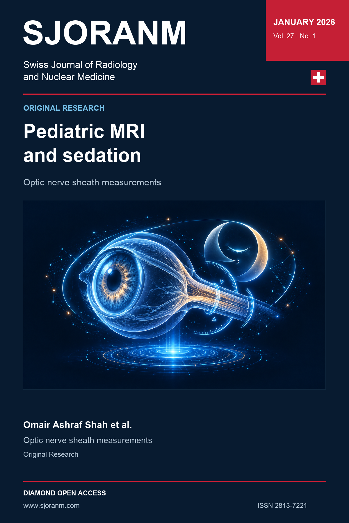
Is Bigger Always Bad ? Evaluating Effect of Sedation on MRI-Based Optic Nerve Sheath Diameter Measurements in Children
DOI:
https://doi.org/10.59667/sjoranm.v27i1.22Keywords:
Intracranial pressure, MRI, Sedation, Optic nerve sheath, ChildrenAbstract
Objectives: To assess the association between sedation status and optic nerve sheath diameter (ONSD) as measured by MRI in pediatric patients.
Materials and Methods: This retrospective observational study included brain MRI examinations of patients aged 0–18 years. Bilateral ONSD was measured on axial T2-weighted images 3 mm posterior to the globe. Results are summarized descriptively by sedation status, and inter-observer agreement is reported using the intraclass correlation coefficient.
Results: Among 78 pediatric patients, 38 underwent MRI with sedation and 40 without sedation. Mean ONSD was 4.8 ± 0.3 mm in the non-sedated group and 5.7 ± 0.4 mm in the sedated group. Inter-observer agreement was reported as an ICC of 0.93 (95% CI: 0.87–0.97).
Conclusion: Higher mean MRI-based ONSD was observed in the sedated group. Sedation status should be considered when interpreting ONSD in children. Prospective studies with appropriate control of confounding factors and independent ICP assessment are needed to determine whether sedation itself affects ONSD and its use as a surrogate for ICP.
References
1. Rajajee V, Vanaman M, Fletcher JJ, Jacobs TL. Optic nerve ultrasound for the detection of raised intracranial pressure. Neurocrit Care 2011;15(3):506-515. https://doi.org/10.1007/s12028-011-9606-8
2. Koziarz A, Sne N, Kegel F, et al. Bedside optic nerve ultrasonography for diagnosing increased intracranial pressure: A systematic review and meta-analysis. Ann Intern Med 2019;171(12):896-905. https://doi.org/10.7326/M19-0812
3. D’Antona L, Asif H, Craven CL, et al. Brain MRI and ophthalmic biomarkers of intracranial pressure. Neurology 2021;96(22):e2714-e2723. https://doi.org/10.1212/WNL.0000000000012023
4. Sallam A, Abdelaal Ahmed Mahmoud MAA, Kamel MG, et al. The diagnostic accuracy of noninvasive methods to measure the intracranial pressure: A systematic review and meta-analysis. Anesth Analg 2021;132(3):686-695. https://doi.org/10.1213/ANE.0000000000005189
5. Gallagher A, Pike M, Berg S. Beware CSF pressure measured under general anaesthesia. Arch Dis Child 2004;89(7):691. https://doi.org/10.1136/adc.2003.032177
6. Kim Y, Choi S, Kang S, Park B. Propofol affects optic nerve sheath diameter less than Sevoflurane during robotic surgery in the steep Trendelenburg position. Biomed Res Int 2019;2019:5617815. https://doi.org/10.1155/2019/5617815
7. Geeraerts T, Newcombe VF, Coles JP, et al. Use of T2-weighted magnetic resonance imaging of the optic nerve sheath to detect raised intracranial pressure. Crit Care 2008;12(5):R114. https://doi.org/10.1186/cc7006
8. Onder H, Goksungur G, Eliacik S, Ulusoy EK, Arslan G. The significance of ONSD, ONSD/ETD ratio, and other neuroimaging parameters in idiopathic intracranial hypertension. Neurol Res 2021;43(12):1098-1106. https://doi.org/10.1080/01616412.2021.1949688
9. Blaivas M, Theodoro D, Sierzenski PR. Elevated intracranial pressure detected by bedside emergency ultrasonography of the optic nerve sheath. Acad Emerg Med 2003;10(4):376-381. https://doi.org/10.1111/j.1553-2712.2003.tb01352.x
10. Maissan IM, Dirven PJ, Haitsma IK, Hoeks SE, Gommers D, Stolker RJ.Ultrasonographic measured optic nerve sheath diameter as an accurate and quick monitor for changes in intracranial pressure. J Neurosurg 2015;123(3):743-747. https://doi.org/10.3171/2014.10.JNS141197
11. Cardim D, Czosnyka M, Chandrapatham K, Badenes R, Bertuccio A, Noto AD, Donnelly J, Pelosi P, Ball L, Hutchinson PJ, Robba C. Effects of Age and Sex on Optic Nerve Sheath Diameter in Healthy Volunteers and Patients With Traumatic Brain Injury. Front Neurol. 2020 Aug 7;11:764. PMID: 32849220; PMCID: PMC7426488. https://doi.org/10.3389/fneur.2020.00764
Downloads
Published
Data Availability Statement
The data that support the findings of this study are available from the corresponding author upon reasonable request. The data contain potentially identifiable patient information and are therefore not publicly available in accordance with Institutional review board and data protection regulations.
Issue
Section
License
Copyright (c) 2026 Omair Ashraf Shah, Mudhabir Ashraf, Akib Arfee, Sumaira Maqbool

This work is licensed under a Creative Commons Attribution 4.0 International License.
This license requires that reusers give credit to the creator. It allows reusers to distribute, remix, adapt, and build upon the material in any medium or format, even for commercial purposes.
 LinkedIn ↗
 X ↗
 VK ↗
 Truth Social ↗
 Bluesky ↗
 YouTube ↗