Vol. 24 No. 1 (2025): Vol. 24 No. 1 (2025)
USA - Eccentric Case of Thoracic Spinal Hemangioma Masquerading as Cauda Equina Syndrome
https://doi.org/10.59667/sjoranm.v24i1.14
We present a case of spinal hemangioma with clinical presentation with lower limb weakness, loss of sensation, back pain, urinary retention and difficulty in bowel movements for 9 months. Patients presenting with lumbar myeloradiculopathy should be carefully evaluated to distinguish it from cauda equina syndrome, spinal dural fistula, spinal cord tumors, and metastasis. Evaluation including MRI brain and CT scan lumbar spine were within normal limits. Further workup including CT scan and MRI thoracic spine revealed T5 vertebral body spinal hemangioma with spina cord compression at T5-T6. The main pathology here is slowly progressive tumor that had power to scrunch the spinal cord and shatter the vertebra, thereby ushering the onset of myeloradiculopathy. Neurosurgery was consulted and they decided to perform a preoperative embolization, and surgical decompression. Recurrence of symptoms despite surgical intervention might necessitate administration of radiation therapy. The neuronal recovery following surgery might be prolonged given the degree of spinal cord injury.
--------------------------------------------------------------------------------------------------------------------------------------------------------
USA - Sporadic Case of LGI1 Autoimmune Encephalitis Feigning as Basal Ganglia Stroke: Exhuming its Pathophysiologic Basis
https://doi.org/10.59667/sjoranm.v24i1.16
LGI1 encephalitis is an immune mediated neurological disorder effecting temporal lobe and basal ganglia. The classic clinical syndrome comprises of myoclonic jerks of the ipsilateral upper arms, facial twitching, memory disturbances, behavioral disturbances and hyponatremia. With its overarching symptoms, it does not snug perfectly with either seizure or movement disorder. The main pathological basis is mass production of anti-LGI1 auto-antibodies which strike the LGI1 protein in the neuronal milieu and presynaptic membrane. On grounds of this immune mediated onslaught, LGI1 protein injury transpires and slowing of signal conduction at the synaptic interface never ensues. In patients with aforementioned symptoms, a high degree of clinical suspicion necessitate MRI brain imaging which shows T2 hyperintensity in basal ganglia, temporal lobe and frontal lobe. This can be corroborated with assessing LGI1 antibody levels in the blood and CSF to monitor disease progression as well as treatment response. As soon as diagnosis is confirmed, intravenous corticosteroids, intravenous immunoglobulins or plasma exchange will reverse the epileptic activity and cognitive dysfunction. Long term immunosuppressive therapy is warranted as it is inclined to reduce the onset of temporal lobe epilepsy and hippocampal atrophy. Regular follow up to detect new clinical symptoms and brain lesions is recommended in these patients to reduce morbidity and mortality.
--------------------------------------------------------------------------------------------------------------------------------------------------------
Argentina - Importance of Molecular Imaging Characteristics for the Diagnosis and Management of Tumor-induced Osteomalacia
https://doi.org/10.59667/sjoranm.v24i1.18
Objective: Tumor-induced osteomalacia (TIO) is a paraneoplastic syndrome associated with the overpro-duction of fibroblast growth factor 23 secondary to phosphaturic mesenchymal tumors (PMT). Our goal was to describe the morphometabolic characterization and histopathological correlation of images obtained from patients with suspected TIO using gallium-68 (68Ga) 1,4,7,10-tetraazacyclodo-decane-1,4,7,10-tetraacetic acid (DOTA)-octreotate (68GaDOTATATE) positron emission tomography /computed tomography (PET/CT) in a referral center in Argentina.
Methods: A prospective, descriptive study with patients suspected of TIO who were referred to confirm the presence of primary lesions by 68Ga-DOTATATE PET/CT.
Results: Eighteen patients were included (female: 72.22%; median age: 47.5 years [range: 41.5–54]). The median maximum standardized uptake value (SUVmax) was 17.2 (interquartile range: 6.27–30.6). Lesions diagnosed by 68Ga-DOTATATE PET/CT were predominantly localized in the appendicular skeleton. Most patients had one identifiable lesion. Lesions were focal and well-limited in 66.67% of cases. Histopathological data were available for 13 patients. PMT was diagnosed in 61.54% of cases; in this subgroup, 25% had lesions showing ill-defined borders and confirmed bone erosion. A numerically, non-significant higher SUVmax was found in patients with PMT. Also, a trend towards isolated soft tissue involvement was more commonly observed among these patients.
Conclusion: In our patients with suspected TIO evaluated by 68Ga-DOTATATE PET/CT, a greater number of lesions were unique, well-defined, and localized in the appendicular skeleton. Nevertheless, ill-defined borders, including bone erosion, were reported in 25% of patients with confirmed PMTs. 68GaDOTATATE PET/CT is a valuable diagnostic tool for patients with suspected TIO. Further research is warranted.
--------------------------------------------------------------------------------------------------------------------------------------------------------
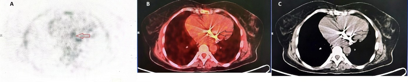
Morocco - Contribution of [18F] FDG PET/CT in the Diagnosis of Prosthetic Valve Infective Endocarditis
https://doi.org/10.59667/sjoranm.v24i1.20
Introduction: Infective endocarditis is a potentially life-threatening disease that requires rapid and accurate diagnosis. Recently, [18F] FDG PET/CT has been incorporated as a new major criterion for the diagnosis of infective endocarditis.
Case report: This report describes a 54-year-old female patient who underwent mitral valve replacement in 2018 for symptomatic severe mitral stenosis with atrial fibrillation. She presented with infectious symptoms on clinical examination. Following transthoracic and transesophageal echocardiography, the differential diagnosis included prosthetic valve infective endocarditis versus mechanical prosthesis thrombosis. [18F] FDG PET/CT ultimately confirmed the diagnosis of prosthetic valve infective endocarditis.
Conclusion: This case highlights the significant advantages of [18F] FDG PET/CT in the diagnostic evaluation of patients with suspected prosthetic valve infective endocarditis, particularly when conventional echocardiographic examinations are inconclusive.