
The Intraosseous Schwannoma of the Upper Extremity
A Single Institutional Experience and Review of Literature
DOI:
https://doi.org/10.59667/sjoranm.v9i1.18Keywords:
Intra-osseous Schwannoma, IOS, Upper Extremity, Upper Limb Tumors, Schwannoma of BonesAbstract
Introduction
Schwannomas are benign soft tissue tumors of neural origin, predominantly occurring in the head and neck regions due to their rich innervation. Intraosseous schwannoma (IOS) is an exceedingly rare form of schwannoma. This study aims to enhance the understanding of intraosseous schwannoma by reviewing cases affecting the upper extremity bones and providing a detailed analysis of their radiographic and magnetic resonance imaging (MRI) characteristics along with a review of the literature on these rare tumors.
Material and Methods
A total of three patients with IOS in upper extremity bones were identified and analyzed. Radiographs and MRI scans were available for all patients. A comprehensive literature review was conducted, including case reports, retrospective studies, and reviews of published data. The epidemiology, anatomical distribution, radiographic characteristics, histological findings, and therapeutic outcomes of intraosseous schwannoma were all investigated, identifying 31 documented cases of IOS involving extremity bones.
Results
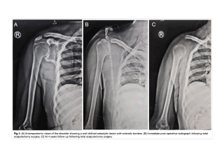
Intraosseous schwannomas primarily affect the mandible, followed by the sacrum and vertebrae. Patients frequently present with non-specific symptoms such as localized pain, swelling, or neurological impairments, which can lead to delayed diagnosis. Radiographic evaluation of IOS typically reveals lytic lesions with well-defined, expansile features and thin sclerotic rims. MRI findings showed that IOS lesions appeared low to iso-intense on T1-weighted images and hyper-intense on T2-weighted images. These imaging characteristics are crucial for differentiating IOS from other lytic bone lesions. Histologically, the presence of Antoni A and Antoni B patterns, as well as S-100 protein positivity, confirms the diagnosis. Surgical management, consisting of curettage, provides a favorable prognosis and low recurrence rates.
Conclusion
Despite its rarity, intraosseous schwannoma should be considered in the differential diagnosis of well-defined, expansile lytic bone lesions, particularly those with thin sclerotic rims. This review provides the most comprehensive analysis to date of IOS affecting extremity bones, emphasizing the importance of recognizing this entity in clinical practice.
References
Wirth WA, Bray CB. Intra-osseous neurilemoma. J Bone Joint Surg. 1977;59A:252–255.
Al-Lhedan F. Schwannoma of the femur: a rare case report. J Bone Oncol. 2017 Sep;1(8):1–3.
Drumond GC, Nakagawa SA, Costa FD, Souza MY, Comunello J, Chung WT. Schwannoma intraosseo: relato de caso e revisão da l i t e r a t u r a . R e v B r a s O r t o p . 2 0 2 0 May;55(15):258–262.
Summers S, Jose J, Barrera CM, Pretell-Mazzini J, Subhawong T, Nguyen NV, Kerr D, Nielsen GP, Rosenberg AE. Intraosseous schwannomas involving the sacrum: Characteristic imaging findings and review of the literature. Neuroradiol J. 2018;31:531–540.
Samter TG, Vellios F, Shafer WG. Neurilemmona of bone. Report of 3 cases with a review of the literature. Radiology. 1960;75:215–222.
Haberal B, Turkbey Simsek D, Simsek EK. Intraosseous schwannoma of the calcaneus: a rare tumor of the bone. Case Rep Orthop. 2018 Oct;2018.
Huajun J, Wei Q, Yuxuan W, Jingjing Y. Intraosseous schwannoma of the proximal humerus with pathologic fracture. Eur J Med Res. 2021 Dec;26(1):1–8.
Meyer A, Billings SD. What's new in nerve sheath tumors. Virchows Arch. 2019;476:65–80.
Ida CM, Scheithauer BW, Yapicier O, Carney JA, Wenger DE, Inwards CY, Bertoni F, Spinner RJ, Unni KK. Primary schwannoma of the bone: a clinicopathologic and radiologic study of 17 cases. Am J Surg Pathol. 2011;35:989–997.
Sun N, Khan UZ, Zeng L, Wu P, Xiong Q, Peng L, Yu H, Tang J. Successful management of a rare radius schwannoma mimicking malignant bone tumors: A case report and literature review. Front Surg. 2023;10:1108942.
Salunkhe R, Limaye S, Biswas SK, Mehta RP. A rare case of calcaneal intraosseous schwannoma. Med J DY Patil Univ. 2012;5:76–78.
Hansra SS, Brown CN, Kang LH, Schaberg KB, Thorpe SW, Chen DC. Partially intraosseous schwannoma of the distal humerus with
increased enhancement after biopsy: Radiologic-pathologic correlation. Radiol Case Rep. 2022 Feb;17(4):1194–1200.
Wahyudi M, Clevfirstarachma RP, Djailani M. Intra-osseous schwannoma of distal femur: A case report. Int J Surg Case Rep. 2022 Oct;99:107643.
Reyniers P, Wafa H, Sinnaeve F, Debeer P, Sciot R. Intraosseous schwannoma of the glenoid: Case report and literature review. SICOT-J. 2021;7:2.
Kamath J, Shetty HB, Madegowda A, Bhatt AS. Intraosseous schwannoma of the humerus: A rarity yet warrants consideration. BMJ Case Rep. 2021;14:e240007.
Lim KX, Wu K. First-ever intraosseous ancient schwannoma of the proximal ulna successfully treated using the cement technique. J Int Med Res. 2021;49:300060520987732.
Mardi K, Vijayamohanan L, Aggarwal V, Kore V. Intraosseous schwannoma of tibia: Report of a rare case with review of literature. Int J Orthop Surg. 2021;29:64.
McAleese T, Clesham K, Moloney D, Hughes A, Faheem N, Merghani K. Intraosseous schwannoma of the femur in a patient with monoclonal gammopathy of undetermined significance. Int J Surg Case Rep. 2020;72:494–498.
Ali SM, Aftab K, Habib Ul Hassan S, Anwar Jilani SA. Intraosseous Schwannoma of fibula: A case report. J Pak Med Assoc. 2022 Jul;72(7):1432–1434.
Nguyen K, Nguyen B. Multi-imaging modalities of intraosseous schwannoma of the scapula. Jt Bone Spine. 2017;84:493.
Gurkan V, Sonmez C, Aralasmak A, Yildiz F, Erdogan O. An Unusual Localization of Intraosseous Schwannoma: The Hamate Bone. Clin Pract. 2017;7:920.
Kim HY, Ryu KN, Park YK, Han JS, Park JS. An Intraosseous Schwannoma Combined with a Subchondral Fracture of the Femoral Head: a Case Report and Literature Review. Investig Magn Reson Imaging. 2017 Sep;21(3):177–182.
Perera N, de Silva C, Perera V. Large schwannoma of the femur – a common tumor at an unusual site: a case report and review of the literature. J Med Case Reports. 2017;11:147.
Suzuki K, Yasuda T, Watanabe K, Kanamori M, Kimura T. Association between intraosseous schwannoma occurrence and the position of the intraosseous nutrient vessel: A case report. Oncol Lett. 2016;11:3185–3188.
Wang G, Wen X, Qu L, Qi X, Yang C. Intraosseous Schwannoma Involving Multiple Bones of the Foot: A Case Report. J Foot Ankle Surg. 2016;55(1):201–206.
Tian YW, Zhang LY, Liu ZQ. Giant intraosseous schwannoma of the scapula: A rare case report and review of the literature. Diagn Pathol. 2014;9:31.
Kito M, Yoshimura Y, Isobe K, Aoki K, Momose T, Kato H. Intraosseous neurilemmoma of the proximal ulna. Int J Surg Case Rep. 2014;5:914–918.
Wang XJ, Hartley K, Holt GE, Fadare O, Cates JM. Intracortical schwannoma of the femur. Skeletal Radiol. 2013;43:687–691.
Wang T, Giugale JM, Ding M, Goodman MA, Schoedel K, Rao UN. Primary intraosseous schwannoma in tibial epiphysis with unique immunohistochemical phenotype: A case report. Int J Surg Pathol. 2014;22:574–578.
Ansari MT, Rastogi S, Alam Khan S, Yadav C, Rijal L. Giant Schwannoma of the First Metatarsal: A Rare Entity. J Foot Ankle Surg. 2014;53:335–339.
Flores Santos F, Pinheiro M, Felicíssimo P. Large foot schwannoma with bone invasion — A case report. Foot Ankle Surg. 2014;20:e23–e26.
Kashima TG, Gibbons MRJP, Whitwell D, Gibbons CLMH, Bradley KM, Ostlere SJ, Athanasou NA. Intraosseous schwannoma in schwannomatosis. Skeletal Radiol. 2013;42:1665–1671.
Hoshi M, Takada J, Oebisu N, Nakamura H. Intraosseous schwannoma of the proximal femur. Asia-Pac J Clin Oncol. 2012;8:e29–e33.
Afshar A, Afaghi F. Intraosseous Schwannoma of the Second Metacarpal: Case Report. J Hand Surg. 2010;35:776–779.
Baˇgci P, Dervi, Soˇglu S, Hiz M, Kanberoˇglu K. Intraosseous schwannoma of the radius. Turk Patoloji Derg/Turk J Pathol. 2010;26:173-176.
Sonig A, Gandhi V, Nanda A. From the cell of Schwann to schwannoma – a century’s fruition. World Neurosurg. 2014;82(5):906–11.
Knight DM, Birch R, Pringle J. Benign solitary schwannomas: a review of 234 cases. J Bone Joint Surg Br. 2007;89:382–387.
Meyer A, Sailhan F, Coulomb A, et al. Proximal tibial epiphyseal intraosseous schwannoma: a rare entity. J Pediatr Orthop. 2008;28(7):786–790.
Jee WH, Oh SN, McCauley T, Ryu KN, Suh JS, Lee JH, Park JM, Chun KA, Sung MS, Kim K, et al. Extraaxial Neurofibromas Versus Neurilemmomas: Discrimination with MRI. Am J Roentgenol. 2004;183:629–633.
Wang YQ, Hu JX, Yang SM, Jiang L, Liu XG, Yuan HS, Wei F, Liu ZJ. Intraosseous schwannoma of the mobile spine: A report of twenty cases. Eur Spine J. 2018;27:3092–3104.
Wirth WA, Bray CB. Intra-osseous neurilemoma: Case report and review of thirty-one cases from the literature. J Bone Joint Surg. 1977;59:252–255.
Downloads
Published
Issue
Section
License
Copyright (c) 2024 ABHIJEET ASHOK SALUNKE

This work is licensed under a Creative Commons Attribution 4.0 International License.
This license requires that reusers give credit to the creator. It allows reusers to distribute, remix, adapt, and build upon the material in any medium or format, even for commercial purposes.


