Vol. 20 No. 1 (2025): Vol. 20 No. 1 (2025)
Case Report:
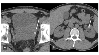
Prostatic Sarcoma
https://doi.org/10.59667/sjoranm.v20i1.16
(from Department of Diagnostic and Interventional Radiology, University Medicine Oldenburg, Carl von Ossietzky Universität Oldenburg, Oldenburg, Germany)
Prostatic sarcomas are rare and aggressive malignancies, often presenting with nonspecific symptoms that mimic benign urological conditions, potentially leading to diagnostic delays [1]. This case outlines the clinical course of a 42-year-old male with a prostatic sarcoma, emphasizing the importance of early imaging, histopathological assessment, and multidisciplinary oncological management.
----------------------------------------------------------------------
Special Area - Highlights from related disciplines:
Pharmacology & Toxicology - Famous Poisonings in History
https://doi.org/10.59667/sjoranm.v20i1.14
(from Royal Academies of Medicine and Related Sciences of Murcia and Valencia, Spain)
The use of poisons spans human history, serving as tools for war, execution, assassination, revenge, and political control. Ancient texts like the "Rig Veda" mention poisoned weapons, and many civilizations used natural toxins—such as frog skin, snake venom, and plant extracts—for lethal purposes. Mythology reflects deep knowledge of poisons. Medea attempted to poison Theseus with aconite to protect her son’s claim to the throne. Hercules used Hydra’s venom to create deadly arrows. In historical contexts, figures like Socrates were executed with poison—hemlock in his case—which was reserved for elite criminals due to its cost. Classical toxicology began in Ancient Greece and continued through the Roman Empire. During Rome’s imperial era, poisons were commonly used in power struggles. Tiberius’ reign saw suspected poisonings of his potential successors, including Germanicus and Drusus. Caligula ultimately rose to power through such intrigue, killing his rivals. Notable toxicologists include Mateo Orfila, who advanced forensic detection techniques in the 19th century, and Juan Bautista Peset Aleixandre, who developed early devices to detect toxic gases in the blood. Natural poisons were also studied in modern science. Cobra venom contains dozens of toxic proteins, many of which disrupt nerve and muscle function. Aconitine, found in "Aconitum napellus", binds to sodium channels in nerves, keeping them open and causing fatal disruptions in cell signaling. Another plant-based toxin, protoanemonin from buttercups, causes painful spasms and ulcers, giving rise to the term "sardonic smile". In Renaissance and Baroque Europe, poisoners like Locusta in Nero’s Rome and La Voisin in Louis XV’s court gained notoriety for their lethal skills. They supplied aristocrats with toxic mixtures to remove rivals or secure inheritances. One infamous potion, “Aqua Tofana”, was linked to hundreds of deaths, possibly including that of Mozart. Venice’s secretive Council of Ten used poison for state security, relying on anonymous citizen reports and aconite-based poisons. In France, women like the Marquise de Brinvilliers and La Voisin were executed for mass poisonings. These individuals often disguised their poisons as medicine or spiritual remedies, exploiting trust and social status. Through myth, science, and scandal, poisons have left an indelible mark on human history, both as instruments of death and as subjects of fascination and fear.
----------------------------------------------------------------------
Original Research:
MRi Regional Strain Analysis in Patients with Hypertrophic Cardiomyopathy
https://doi.org/10.59667/sjoranm.v20i1.18
(from Bristol Heart Institute, University Hospitals Bristol and Weston NHS Foundation Trust 2025, Bristol, United Kingdom)
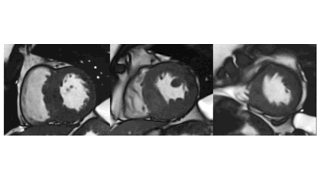
Purpose: To evaluate the regional left ventricular myocardial strain in patient with hypertrophic cardiomyopathy (HCM) especially young apparently compensated patients by magnetic resonance imaging.
Materials and Methods: 25 HCM patients representing all age groups and 25 healthy volunteers underwent 1.5 Tesla MRI examination for cardiac volumes, and mass, followed by regional strain analysis in radial, circumferential, and longitudinal directions as regard the displacement, strain, peak diastolic and systolic strain rate, peak diastolic and systolic velocity, time to peak displacement and time to peak strain.
Results: In the HCM group, hypertrophic segments showing delayed gadolinium enhancement (DGE) were significantly different from non-hypertrophic apparently normal showing no enhancement concerning most of the regional radial strain parameters.
In longitudinal and circumferential directions, hypertrophic segments showing DGE were significantly different from apparently normal segments with no enhancement as regard the strain, peak diastolic and systolic strain rate. Compared to normal volunteers, the hypertrophic segments with DGE were significantly different concerning most of the radial and longitudinal strain parameters, while apparently normal segments with no enhancement don’t present a similar significant difference.
In circumferential analysis, hypertrophic enhancing segments were significantly different as regard the strain, peak diastolic strain rate, peak systolic velocity, and time to peak displacement, while the apparently normal non-enhancing segments present difference concerning the strain, peak systolic velocity, and time to peak displacement.
Conclusion: The hypertrophied segments are more affected than the segments of apparently normal thickness in HCM patients, especially in the radial direction, the apparently normal segments are also affected with no tendency of functional compensation. Our data show that HCM muscle fibers show mostly hypofunction and reduced contraction rather than hypercontraction and even the apparently normal segments are impaired to a certain degree in comparison to normal. These findings are essential in follow up studies especially with patient receiving myosin inhibitors as well as young patients presenting preserved cardiac function and for gene positive apparently normal with absent penetration of the gene in the form of a concentric thickening can be seen by imaging, do we need more sophisticated imaging process like strain analysis to ensure absent penetration.