A Comparison of CT and MR Imaging of Cystic Renal Masses by Using the Bosniak Classification System
DOI:
https://doi.org/10.59667/sjoranm.v4i1.14Keywords:
renal mass, Bosniak classification, comparison of CT and MRI, diagnostic value, intracystic septa, cystic wall thickeningAbstract
Purpose:
To compare the efficacy of computed tomography (CT) and magnetic resonance imaging (MRI) in evaluating cystic renal masses using the Bosniak classification system.
Materials and Methods:
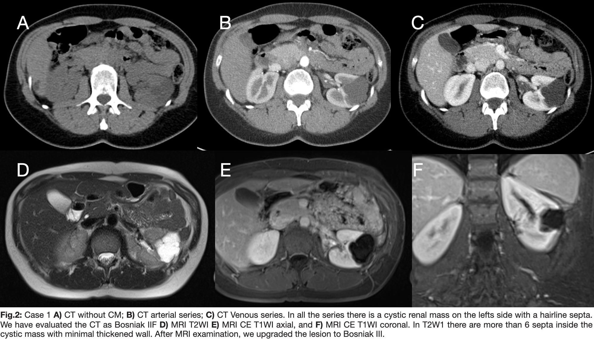
Between January 2010 and December 2020, a total of 844 patients with suspected renal masses underwent total or partial nephrectomy at the Department of Urology, Heidelberg University Clinic. Among them, 123 patients presented with cystic renal masses. Out of this cohort, 15 patients underwent preoperative examinations using both MRI and CT within a 6-week timeframe. These examinations were retrospectively analyzed by two radiologists employing the Bosniak classification system. Each lesion was assessed based on CT images for lesion localization, volume, number, and thickness of septa/wall, calcification, presence of enhancing soft-tissue components, and lesion density. Pathologic correlation was available for all lesions.
Results:
Out of the 844 patients, 15 underwent both CT and MR imaging examinations (male-to-female ratio: 2.75, age range: 43-75 years; mean age: 60.5 years). Based on CT images, there were one category I, one category II, three category IIF, three category III, and seven category IV lesions. Findings based on CT and MR images were concordant in eleven (73.3%) renal lesions, while in 4 (26.7%) masses, differences were observed. In three (20%) cases, MR imaging revealed more septa/wall than CT, resulting in an upgrade of the classification at MR imaging. In one (6.7%) lesion, MR imaging showed no wall and/or septa thickness compared with CT, leading to a classification downgrade. MR imaging results led to a cyst classification upgrade in three masses (from category II to III, n=1; IIF to III, n=2) and downgrade in one lesion (from category IIF to II). Pathologically, 11 lesions (73.3%) were malignant (9 Cystic Clear cell carcinoma and 2 papillary renal cell carcinoma), while 4 lesions were renal cysts without malignancy.
Conclusion:
The majority of findings from both CT and MR imaging were similar in suspected cystic renal masses. MR imaging demonstrated additional septa/wall or septa/wall thickening, potentially upgrading lesions from borderline category IIF to III compared to CT. This distinction may contribute to improved case management.
References
Brant WA, Helms CA (2007); Fundamentals of Diagnostic Radiology, 879 – 885; In: Genitournary, Adrenal Glands and Kidneys. Second edition. Lippincott Williams & Wilkins, New York
Slone RM et al. (1999); Body CT: A Practical Approach; In: Kidney. Edition. McGraw-Hill Publishing Co., New York
Isreal GM, Bosniak MA (2005); An update of the Bosniak renal cyst classification system. Radiology 236:441–450
Bosniak MA (1986); The current radiological approach to renal cysts. Radiology 158:1–10
Siegel CL (1997); CT of cystic renal masses: analysis of diagnostic performance and interobserver variation. AJR Am J Roentgenol 169:813– 818
Bosniak MA (2004); Evaluation of Cystic Renal Masses: Comparison of CT and MR Imaging by Using the Bosniak Classification System. Radiology 231:365–371
Israel GM, Bosniak MA (2005); How I Do It: Evaluating Renal Masses. Radiology 236:441–450
Blandino et al. (2002); MR urography of the ureter. AJR Am J Roentgenol 179:1307-1314
Israel GM, Bosniak MA (2005); Pitfalls in Renal Mass Evaluation and How to Avoid Them; Radio-Graphics 2008; 28:1325–1338
Bosniak MA (2012); The Bosniak Renal Cyst Classification: 25 Years Later; Radiology 262: 781 –785
Balci NC et al. (1999); Complex renal cysts: findings on MR imaging; AJR Am J Roentgenol 172:1495–1500.
Adey GS et al. (2008); Lower limits of detection using magnetic resonance imaging for solid components in cystic renal neoplasms; Urology 71:47–51
Eble JN et al. (2004); Tumors of the kidney in: WHO classification of tumors: tumors of the urinary system and male genital organs IARC Press, Lyon, France
Hecht EM et al. (2004); Renal masses: quantitative analysis of enhancement with signal intensity measurements versus qualitative analysis of enhancement with image subtraction for diagnosing malignancy at MR imaging; Radiology 232:373–378
Inci E et al. (2012); Diffusion-weighted magnetic resonance imaging in evaluation of primary solid and cystic renal masses using the Bosniak classification; Eur J Radiol. 81: 815-20.
Squillaci E et al. (2004); Diffusion-weighted MR imaging in the evaluation of renal tumours; J Exp Clin Cancer Res. 23: 39-45.
Roussel, E., Capitanio, U., Kutikov, A., Oosterwijk, E., Pedrosa, I., Rowe, S. P., & Gorin, M. A. (2022). Novel imaging methods for renal mass characteri-zation: a collaborative review. European Uro-logy, 81(5), 476-488.
Klaus, J., Huber, D. A., Daneshvar, K., Prosch, H., Christe, A., & Ebner, L. (2018). Renale Hämosiderose durch intravaskuläre Hämolyse. RoFo: Fortschritte auf dem Gebiete der Röntgenstrahlen und der Nuklearmedizin, 190(11), 1072–1073. https://doi.org/10.1055/a-0626-7534
Huber, J., Winkler, A., Jakobi, H., Bruckner, T., Roth, W., Hallscheidt, P., Daneshvar, K., Hohenfellner, M., & Pahernik, S. (2014). Preoperative decision making for renal cell carcinoma: cystic morphology in cross-sectional imaging might predict lower malignant potential. Urologic oncology, 32(1), 37.e1–37.e376.

Downloads
Published
Issue
Section
License
Copyright (c) 2024 Keivan Daneshvar, Johannes Huber, Wolfram Andreas Bosbach, Nando Mertineit, Gerd Nöldge

This work is licensed under a Creative Commons Attribution 4.0 International License.
This license requires that reusers give credit to the creator. It allows reusers to distribute, remix, adapt, and build upon the material in any medium or format, even for commercial purposes.


