Vol. 9 No. 1 (2024): Vol. 09 No. 1 (2024)
Primary extrauterine choriocarcinoma is a rare condition, commonly reported in the cervix. Primary vulvovaginal choriocarcinoma has rarely been reported and review of literature reveals no case with MRI features for choriocarcinoma involving only the vulva. We report a rare case of primary choriocarcinoma of the vulva in a young woman, highlighting its Magnetic Resonance Imaging (MRI) features.
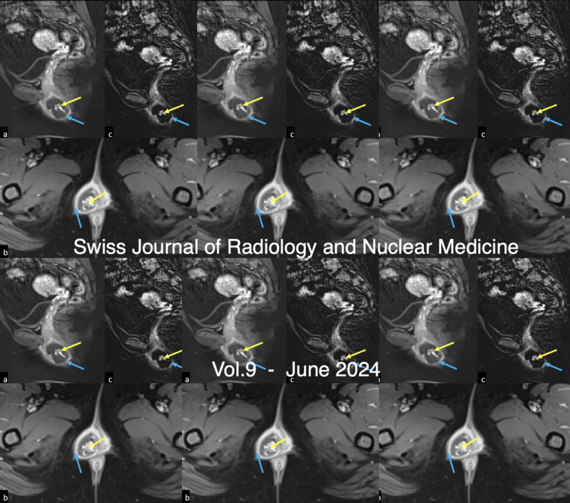
A 26-year-old female patient presented with complaints of menorrhagia, elevated serum Beta human chorionic gonadotropin (β-hCG) levels and a history of suction evacuation for partial molar pregnancy 4 months ago. Magnetic Resonance Imaging (MRI) of the pelvis revealed a normal uterus and adnexa. However, there was a well-defined T1 hypointense, T2 heterogenously hyperintense sublabial mass lesion with internal flow voids, few peripheral areas of restricted diffusion and few internal areas of T1 hyperintensity that bloomed on gradient images. On contrast administration, there was intense peripheral enhancement of the lesion, especially in the regions of restricted diffusion and also in the center of the lesion, where flow voids were observed. Few confluent non enhancing areas of necrosis were also present. In view of the clinical presentation, elevated β-hCG and imaging findings, a diagnosis of choriocarcinoma of the vulva was made and chemotherapy (EMA-CO regimen) was initiated, following which β-hCG decreased progressively and the patient is currently asymptomatic and follow up MRI revealed no abnormal significant residual lesion.
Awareness of a labial lesion being due to extrauterine choriocarcinoma in patients with elevated β-hCG is crucial for effective management and recovery. Our case uniquely emphasizes pretreatment MRI features as well as post-treatment imaging follow-up.
--------------------------------------------------
The Utility of multimodality imaging in a curious case of crippled child
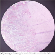
The aim of the article is to establish the importance of devising and adhering to an imaging protocol for musculoskeletal imaging by presenting a case of Dysplasia epiphysealis hemimelica also known as Trevor’s disease in a 13 year old child with atypical presentation which proved to be a diagnostic challenge.