
The MRI regional strain analysis in patients with hypertrophic cardiomyopathy
DOI:
https://doi.org/10.59667/sjoranm.v20i1.18Keywords:
Strain MRI, Hypertrophic cardiomyopathy, Magnetic Resonance (MRI)Abstract
Purpose: To evaluate the regional left ventricular myocardial strain in patient with hypertrophic cardiomyopathy (HCM) especially young apparently compensated patients by magnetic resonance imaging.
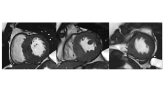
Materials and Methods: 25 HCM patients representing all age groups and 25 healthy volunteers underwent 1.5 Tesla MRI examination for cardiac volumes, and mass, followed by regional strain analysis in radial, circumferential, and longitudinal directions as regard the displacement, strain, peak diastolic and systolic strain rate, peak diastolic and systolic velocity, time to peak displacement and time to peak strain.
Results: In the HCM group, hypertrophic segments showing delayed gadolinium enhancement (DGE) were significantly different from non-hypertrophic apparently normal showing no enhancement concerning most of the regional radial strain parameters.
In longitudinal and circumferential directions, hypertrophic segments showing DGE were significantly different from apparently normal segments with no enhancement as regard the strain, peak diastolic and systolic strain rate.
Compared to normal volunteers, the hypertrophic segments with DGE were significantly different concerning most of the radial and longitudinal strain parameters, while apparently normal segments with no enhancement don’t present a similar significant difference.
In circumferential analysis, hypertrophic enhancing segments were significantly different as regard the strain, peak diastolic strain rate, peak systolic velocity, and time to peak displacement, while the apparently normal non-enhancing segments present difference concerning the strain, peak systolic velocity, and time to peak displacement.
Conclusion
The hypertrophied segments are more affected than the segments of apparently normal thickness in HCM patients, especially in the radial direction, the apparently normal segments are also affected with no tendency of functional compensation.
Our data show that HCM muscle fibers show mostly hypofunction and reduced contraction rather than hypercontraction and even the apparently normal segments are impaired to a certain degree in comparison to normal.
These findings are essential in follow up studies especially with patient receiving myosin inhibitors as well as young patients presenting preserved cardiac function and for gene positive apparently normal with absent penetration of the gene in the form of a concentric thickening can be seen by imaging, do we need more sophisticated imaging process like strain analysis to ensure absent penetration.
References
1) Minhas, A. M. K., Wyand, R. A., Ariss, R. W., Nazir, S., Shahzeb Khan, M., Jia, X., Greene, S. J., Fudim, M., Wang, A., Warraich, H. J., Kalra, A., Alam, M., & Virani, S. S. (2022). Demographic and Regional Trends of Hypertrophic Cardiomyopathy-Related Mortality in the United States, 1999 to 2019. Circulation: Heart Failure, 15(9), E009292. https://doi.org/10.1161/CIRCHEARTFAILURE.121.009292.
2) Semsarian C, Ingles J, Maron MS, Maron BJ. New perspectives on the prevalence of hypertrophic cardiomyopathy. J Am Coll Cardiol (2015); 65:1249–1254. https://www.jacc.org/doi/full/10.1016/j.jacc.2015.01.019
3) Baxi AJ, Restrepo CS, Vargas D, et al. Hypertrophic cardiomyopathy from A to Z: Genetics, pathophysiology, imaging, and management. Radiographics (2016);36(2):335-54. https://doi.org/10.1148/rg.2016150137
4) Amano, Y., Kitamura, M., Takano, H., Yanagisawa, F., Tachi, M., Suzuki, Y., Kumita, S., & Takayama, M. (2018). Cardiac MR imaging of hypertrophic cardiomyopathy: Techniques, findings, and clinical relevance. In Magnetic Resonance in Medical Sciences (Vol. 17, Issue 2, pp. 120–131). Japanese Society for Magnetic Resonance in Medicine. https://doi.org/10.2463/mrms.rev.2017-0145
5) Cui, H., Schaff, H. v., Lentz Carvalho, J., Nishimura, R. A., Geske, J. B., Dearani, J. A., Lahr, B. D., Lee, A. T., Bos, J. M., Ackerman, M. J., Ommen, S. R., & Maleszewski, J. J. (2021). Myocardial Histopathology in Patients With Obstructive Hypertrophic Cardiomyopathy. Journal of the American College of Cardiology, 77(17), 2159–2170. https://doi.org/10.1016/j.jacc.2021.03.008
6) Almaas, V., and Amlie, P. Histopathological Changes and Clinical Implications in Patients with Hypertrophic Cardiomyopathy, European Cardiology (2010);6(2):88–90 https://doi.org/10.15420/ecr.2010.6.2.88
7) James A. Spudich J. Three perspectives on the molecular basis of hypercontractility caused by hypertrophic cardiomyopathy mutations. European Journal of Physiology (2019) 471:701–717. https://doi.org/10.1007/s00424-019-02259-2
8) Olivotto, I., Cecchi, F., Poggesi, C., & Yacoub, M. H. (2012). Patterns of disease progression in hypertrophic cardiomyopathy an individualized approach to clinical staging. Circulation: Heart Failure, 5(4), 535–546. https://doi.org/10.1161/CIRCHEARTFAILURE.112.967026
9) Marian, A. J., & Braunwald, E. (2017). Hypertrophic cardiomyopathy: Genetics, pathogenesis, clinical manifestations, diagnosis, and therapy. Circulation Research, 121(7), 749–770. https://doi.org/10.1161/CIRCRESAHA.117.311059
10) Ommen SR, Mital S, Burke MA, Day SM, Deswal A, Elliott P, et al. (2020) AHA/ACC Guideline for the Diagnosis and Treatment of Patients With Hypertrophic Cardiomyopathy: A Report of the American College of Cardiology/American Heart Association Joint Committee on Clinical Practice Guidelines. J Am Coll Cardiol 2020;162:e23-106. https://doi.org/10.1161/CIR.0000000000000937
11) Lopez L, Colan S, Stylianou M, Granger S, Trachtenberg F, Frommelt P, et al. Relationship of Echocardiographic Z Scores Adjusted for Body Surface Area to Age, Sex, Race, and Ethnicity: The Pediatric Heart Network Normal Echocardiogram Database. Circ Cardiovasc Ima-ging (2017);10: e006979. https://doi.org/10.1161/CIRCIMAGING.117.006979
12) Reant P, Reynaud A, Pillois X, et al. Comparison of resting and exercise echocardiographic as indicators of outcomes in hypertrophic. J Am Soc Echocardiogr (2015);28:194–203. https://doi.org/10.1016/j.echo.2014.10.001
13) Reant P, Mirabel M, Lloyd G, et al. Global longitudinal strain is associated with heart failure outcomes in hypertrophic cardiomyopathy. Heart (2016);102:741–747. https://doi.org/10.1136/heartjnl-2015-308576
14) Kawel-Boehm, N., Maceira, A., Valsangiacomo-Buechel, E. R., Vogel-Claussen, J., Turkbey, E. B., Williams, R., Plein, S., Tee, M., Eng, J., & Bluemke, D. A. (2015). Normal values for cardiovascular magnetic resonance in adults and children. In Journal of Cardiovascular Magnetic Resonance (Vol. 17, Issue 1). BioMed Central Ltd. https://doi.org/10.1186/s12968-015-0111-7
15) Kawel-Boehm, N., Hetzel, S. J., Ambale-Venkatesh, B., Captur, G., Francois, C. J., Jerosch-Herold, M., Salerno, M., Teague, S. D., Valsangiacomo-Buechel, E., van der Geest, R. J., & Bluemke, D. A. (2020). Reference ranges (“normal values”) for cardiovascular magnetic resonance (CMR) in adults and children: 2020 update. In Journal of Cardiovascular Magnetic Resonance (Vol. 22, Issue 1). BioMed Central Ltd. https://doi.org/10.1186/s12968-020-00683-3
Downloads
Published
Data Availability Statement
All data generated or analyzed during this study are available from the corresponding author upon reasonable request. The datasets supporting the conclusions of this article have been securely archived and can be accessed for further assessment, subject to ethical and legal considerations. This work has been published in the Swiss Journal of Radiology and Nuclear Medicine.
Issue
Section
License
Copyright (c) 2025 Esraa Maher, Magdi Khalil, Mona Taher, Sameh Khalil

This work is licensed under a Creative Commons Attribution 4.0 International License.
This license requires that reusers give credit to the creator. It allows reusers to distribute, remix, adapt, and build upon the material in any medium or format, even for commercial purposes.


