Vol. 21 No. 1 (2025): Vol. 21 No. 1 (2025)
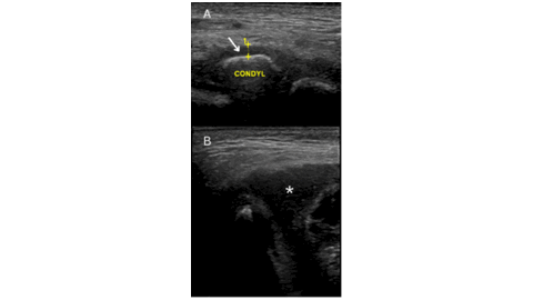
Septic arthritis of the temporomandibular joint in a 6-year-old boy: A case report
https://doi.org/10.59667/sjoranm.v21i1.16
Septic arthritis of the temporomandibular joint (TMJ) is an uncommon condition, particularly in pediatric patients. We present a case of a previously healthy 6-year-old boy who presented with septic arthritis of the TMJ with extra-articular involvement, which responded well to an extended course of intravenous antibiotic therapy, leading to complete resolution on follow-up imaging.
-----------------------------------------------------
Radiology Display Technology: Progress Over Time and the Role of Standards Today
https://doi.org/10.59667/sjoranm.v21i1.28
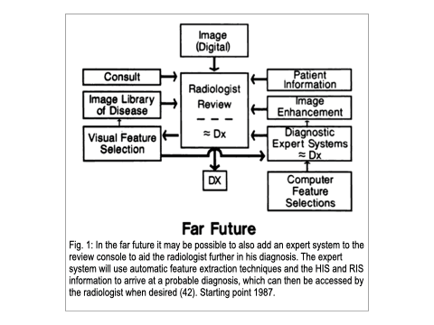
Not long ago, X-ray information was recorded on film. Consequently, after development and fixation, post-processing of the image as we use today was simply only possible through another X-ray exposure with additional radiation and uncertain results. The introduction of the digital image information chain from the X-ray detector to the monitor has fundamentally changed this. The digital transformation of radiology has been continuously expanded and improved through the application of new and increasingly powerful technical components. The omnipresence of radiological image information extends from the place of creation via PACS (Picture Archiving and Communication System) not only within the radiology department but also throughout the entire hospital and its departments, such as emergency room, operating room, wards, and outpatient clinics of the referring specialties. Further dissemination of digital image information occurs via CD, DVD, USB sticks, and via the internet through patient and referrer portals. The end display devices of the image recipients/users can be projectors, beamers, computer screens, tablets, televisions, smartphones, or other electronic devices with suitable displays. In fact, visualizations of X-ray images on not-too-large displays of car radios are conceivable, for example, if a WhatsApp image message arrives via mobile phone to a radiologist driving a car. Following the desires of the regulatory authorities, all these displays would have to be continuously checked for their display quality because it cannot be ruled out that an X-ray image might be displayed. Theoretically, this is conceivable. However, it is simply not feasible in our overregulated reality by now. Fact-based arguments are discussed regarding this issue, covering various aspects of the diagnostic significance and the technical physical specifications of radiological images. Thus, we provide lawmakers and authorities with evidence-based facts to ensure that future legislative measures appropriately regulate radiologic display quality.
-----------------------------------------------------
A Must-Know Guide to Complex Anal Fistulas Supralevator, Extrasphincteric, and More
https://doi.org/10.59667/sjoranm.v21i1.14
Treatment of perianal fistulae, especially high-grade Grade 5 perianal fistula, is still a difficult procedure in colorectal surgery. Imaging is central to the correct classification, diagnosis, and preoperative evaluation of fistulae. The purpose of this article is to briefly present the five types of perianal fistula with Grade 5 complexity according to the radiological aspects, classification, and therapeutic implications.
-----------------------------------------------------
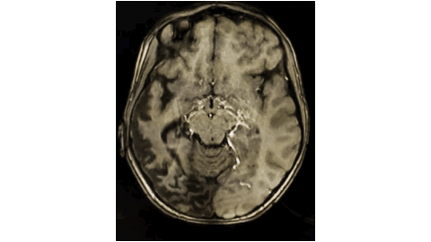
Unravelling paediatric stroke: a rare presentation of paediatric Moya moya disease with simultaneous involvement of anterior and posterior circulation https://doi.org/10.59667/sjoranm.v21i1.20Moya Moya disease is a rare cerebrovascular disorder characterized by progressive stenosis of the internal carotid arteries and their branches, leading to a range of neurological symptoms. This case report discusses a 14-year-old boy presenting with classic symptoms associated with Moya Moya disease. The diagnostic process involved imaging studies, which confirmed the characteristic findings of the disease along with a specific ANA study which further helped in excluding the association of any other autoimmune disease. Treatment options were explored, including, the use of therapeutic drugs like Aspirin and some statins, aimed at preventing TIAs and alleviating symptoms. This case highlights the importance of early recognition and management of Moya Moya disease to prevent serious neurological deficits and improve the quality of life in affected children and adolescents. Further research is needed to explore the long-term outcomes of various treatment strategies for this challenging condition.
-----------------------------------------------------
Sonographic Evaluation of Male Testicular Volume Amongst Patients With Fertility Challenges: A Cross-Sectional Retrospective Study
https://doi.org/10.59667/sjoranm.v21i1.18
Background
Male infertility is a significant health issue affecting approximately 15% of couples worldwide. Testicular volume has been suggested as a potential predictor of fertility issues in men, but no study has correlated testicular volume amongst men with infertility challenges in our environment. The purpose of the study is to evaluate the relationship between testicular volume and male fertility profile in subjects with fertility challenges.
Methods
The research involves a cross-sectional retrospective analysis of records of male patients that presented to the teaching hospital with fertility concerns. Records containing age, Ultrasound measurement of testicular volume, sperm count, and sperm motility were taken into account. Sample size used was 50 records of male patients that presented with infertility. Independent samples t-test and Pearson correlation were employed at a 5% level of significance.
Results
Our results suggest that patients with lower testicular volume may have compromised sperm production and quality, thus contributing to infertility. There was a significant difference between the testicular volume of infertile males and fertile males [RT. Volume (p = <0.0001) & LT. Volume (p < 0.0001)]. Findings in correlating testicular volume with sperm parameters shows there was a significant relationship between sperm motility sluggish and left testicular volume (r = 0.301, p = 0.034) and between sperm count and left testicular volume (r = 0.317, p = 0.025). For sperm motility activeness (r = 0.092, p = 0.532) and sperm motility deadness (r = 0.031, p = 0.828), there was no significant relationship.
Discussion
The result from the analysis showed there was a positive significant difference in the values of the testicular volumes of infertile and fertile male patients with the testicular volume for infertile males lesser than the normative volume for fertile males.
Conclusion
This study showed that the mean testicular volume in male with fertility challenges are lesser. Smaller testicular volume was associated more with lesser sperm motility sluggishness and reduced sperm count. This implies that clinicians could include testicular volume measurements as part of routine fertility assessments to help identify individuals at risk for infertility.
-----------------------------------------------------
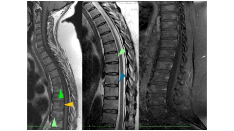
A disconcerting sarcoidosis, when the spine gets involved...
https://doi.org/10.59667/sjoranm.v21i1.22
Bone involvement in sarcoidosis is uncommon and may be discovered incidentally when pain appears in affected patients. Indeed, the pelvic-spinal location is the most widespread: It can manifest when the spine is affected by inflammatory pain, sometimes mimicking authentic spondyloarthritis in certain cases, and may even suggest a malignant lesion by its radiological appearance. We describe the original observation of a patient suffering from inflammatory and insomnia-causing spinal pain in whom the diagnosis of mediastinal sarcoidosis was retained in therapeutic abstention on the pulmonary level.
Observation: 52-year-old female patient who had presented for barely a year a chronic cough accom-panied by basithoracic pain, leading her to perform a chest CT scan which found bilateral mediastinal-hilar adenopathies and peri-lymphatic pulmonary nodules. The diagnosis of type 2 sarcoidosis was immediately considered. The additional investigations have indeed confirmed the suspected disease on the clinical and radiological level, in particular by converse enzyme positivity, the tuberculin anergy on the IDR, the lymphocyte predominance in the bronchoalveolar fluid but more particularly by the demonstration of the epithelial-giant cellular granuloma on the right iliac lymph node biopsy. However, the patient presents with intense inflammatory back pain, sometimes causing insomnia. A spinal MRI showed no-dular bone marrow replacement lesions with pronounced T1 hyposignal and T2 hypersignal enhanced after gadolinium injection. Fortunately, she does not present bone fractures and no osteoporosis on bone densitometry.
Conclusion: Faced with spinal symptoms in a context of sarcoidosis, bone involvement must be considered, especially since there is no correlation between the clinical picture and imaging. The latter most often has a favorable prognosis in the absence of fractures despite sometimes suspicious imaging. The case of our patient clearly illustrates the bone-related nature of the disease; back pain can be particularly intense and should lead to discussion of the use of corticosteroids or even basic treatment for rheumatic diseases.
-----------------------------------------------------
A Radiologist's view on Pedal Actinomycetoma: A Case Report with Comprehensive Review of Literature
https://doi.org/10.59667/sjoranm.v21i1.26
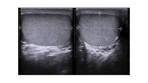
Background: Tropical diseases comprise of an array of communicable and non-communicable diseases that prevail in the tropical belt. Madura foot, classified as a tropical disease by WHO, is a chronic granulomatous disease that predominantly involves the skin and subcutaneous tissue, commonly affecting the lower limbs. We present a case of actinomycetoma with extensive review of the existing literature, focusing on diagnostic imaging.
Case presentation: A 36-year-old female from eastern India presented with a six-month history of right foot swelling and a discharging wound. She was unsuccessfully treated with multiple courses of antibiotics in local hospitals. Upon referral, radiological investigations were performed for further evaluation. USG showed infiltrative hypoechoic soft tissue with nodular lesions showing targetoid appearance. MRI revealed infiltrative soft tissue with variable sized nodular lesion showing characteristic ‘dot-in-circle' appearance, prompting the diagnosis of pedal mycetoma. Actinomycetoma was confirmed on biopsy.
Conclusion: Pedal mycetoma presents significant diagnostic and therapeutic challenges owing to its insidious progression and delayed diagnosis. Radiological imaging, particularly MRI, plays a pivotal role in diagnosis and staging of the disease, enabling detailed evaluation of soft tissue and bone involvement. The ‘dot-in-circle' sign observed on imaging is pathognomic and aids in accurate diagnosis. Early dia-gnosis facilitated by diagnostic imaging warrants improved therapeutic outcomes.