
A Disconcerting Sarcoidosis, When The Spine Gets Involved...
DOI:
https://doi.org/10.59667/sjoranm.v21i1.22Keywords:
bone sarcoidosis, inflammatory back pain, spinal MRIAbstract
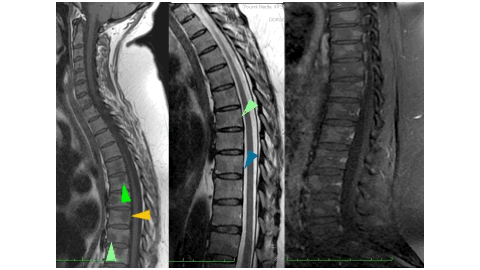
Bone involvement in sarcoidosis is uncommon and may be discovered incidentally when pain appears in affected patients. Indeed, the pelvic-spinal location is the most widespread: It can manifest when spine is affected by inflammatory pain, sometimes mimicking authentic spondylarthritis in certain cases, and may even suggest a malignant lesion by its radiological appearance. We describe the original observation of a patient suffering from inflammatory and insomnia-causing spinal pain in whom the diagnosis of mediastinal sarcoidosis was retained in therapeutic abstention on the pulmonary level. Observation: 52-year-old female patient who had presented for barely a year a chronic cough accompanied by basithoracic pain, leading her to perform a chest CT scan which found bilateral medistinal-hilar adenopathies and peri-lymphatic pulmonary nodules. The diagnosis of type2 sarcoidosis was immediately considered. The additional investigations have indeed confirmed the suspected disease on the clinical and radiological level, in particular y converse enzyme positivity, the tuberculin anergy on the IDR, the lymphocyte predominance in the bronchoalveolar fluid but more particularly by he demonstration of the epithelial-giant cellular granuloma on the right iliac lymph node biopsy. However, the patient presents with inflammatory back pain, sometimes causing insomnia. A spinal MRI showed nodular bone marrow replacement lesions with pronounced T1 hyposignal and T2 hypersignal enhanced after gadolinium injection. Fortunately, she does not present bone fractures and no osteoporosis on bone densitometry. Conclusion: Faced with spinal symptoms in a context of sarcoidosis, bone involvement must be considered, espacially since there is no correlation between the clinical picture and imaging. The latter most often has a favorable prognosis in the absence of fractures despite sometimes suspicious imaging. The case of our patient clearly illustrates the bone-related nature of the disease; back pain can be particularly intense and should lead to discussion of the use of corticisteroids or even basic treatment for rheumatic diseases.
References
1) Wilcox A, Bharadwaj P, Sharma OP. Bone sarcoidosis. Curr Opin Rheumatol 2000; 12:321–30. https://journals.lww.com/co-rheumatology/abstract/2000/07000/bone_sarcoidosis.16.aspx
2) Soussan M, Augier A, Brillet PY, Weinmann P, Valeyre D. Functional imaging in extra-pulmonary sarcoi-dosis: FDG-PET/CT and MR features. Clin Nucl Med 2014; 39: e146–59. https://doi.org/10.1097/RLU.0b013e318279f264
3) Zhou Y, Lower EE, Li H, Farhey Y, Baughmann RP. Clinical characteristics of patients with bone sarcoidosis. Semin Arthritis Rheum 2017 ; 47:143–8. https://doi.org/10.1016/j.semarthrit.2017.02.004
4) Challal S, Rivière E, Lanseur B, et al. Axial bone involvement in sarcoidosis has a good prognosis: longitudinal study of a cohort of 48 patients. Semin Arthritis Rheum 2025; 71:152654. https://doi.org/10.1016/j.semarthrit.2025.152654
5) Ben Hassine I, Rein C, Comarmond C, et al. Osseous sarcoidosis: a multicenter retrospective case-control study of 48 patients. Joint Bone Spine 2019; 86:789–93. https://doi.org/10.1016/j.jbspin.2019.07.009
6) Moore SL, Kransdorf MJ, Schweitzer ME, Murphey MD, Babb JS. Can sarcoidosis and metastatic bone lesions be reliably differentiated on routine MRI? Am J Roentgenol 2012; 198:1387– 93. https://doi.org/10.2214/AJR.11.7498
7) Conte G, Zugni F, Colleoni M, Renne G, Bellomi M, Petralia G. Sarcoidosis with bone involvement mimicking metastatic disease at (18)F-FDG PET/CT: problem solving by diffusion whole-body MRI. Ecancermedicalscience 2015; 9:537.https://doi.org/10.3332/ecancer.2015.537
8) Kobak S, Sever F, Ince O, Orman M. The prevalence of sacroiliitis and spondyloarthritis in patients with sarcoidosis. Int J Rheumatol 2014; 2014:289454. https://doi.org/10.1155/2014/289454
9) Wu C-H, Chung P-I, Wu C-Y, et al. Comorbid autoimmune diseases in patients with sarcoi-dosis: a nationwide case-control study in Taiwan. J Dermatol 2017; 44:423–30. https://doi.org/10.1111/1346-8138.13654
10) Sigaux J, Semerano L, Nasrallah T, et al. High prevalence of spondyloarthritis in sarcoidosis patients with chronic back pain. Semin Arthritis Rheum 2019; 49:246–50. https://doi.org/10.1016/j.semarthrit.2019.03.006
11) Cadiou S, Robin F, Guillin R, et al. Spondyloarthritis and sarcoidosis: related or fake friends? A systematic literature review. Joint Bone Spine 2020; 87:579–87. https://doi.org/10.1016/j.jbspin.2020.06.011
12) Demaria L, Borie R, Benali K, et al. 18F-FDG PET/CT in bone sarcoidosis: an observational study. Clin Rheumatol 2020 ; 39:2727–34. https://doi.org/10.1007/s10067-020-05022-6
13) Challal S, Rivière E, Lanseur B, et al. Axial bone involvement in sarcoidosis has a good prognosis: longitudinal study of a cohort of 48 patients. Semin Arthritis Rheum 2025; 71:152654. https://doi.org/10.1016/j.semarthrit.2025.152654
14) Sparks JA, McSparron JI, Shah N, et al. Osseous sarcoidosis: clinical characteristics, treatment, and outcomes – experience from a large, academic hospital. Semin Arthritis Rheum 2014; 44:371–9. https://doi.org/10.1016/j.semarthrit.2014.07.003
15) Grozdic Milojevic I, Sobic-Saranovic D, Videnovic-Ivanov J, Saranovic D, Odalovic S, Artiko V. FDG PET/CT in bone sarcoidosis. Sarcoidosis Vasc Diffuse Lung Dis 2016; 33:66674. https://pubmed.ncbi.nlm.nih.gov/27055838/
Downloads
Published
Data Availability Statement
Not applicable.
Issue
Section
License
Copyright (c) 2025 Selma Abdellaoui, Prof. Dr. Kamal Allal

This work is licensed under a Creative Commons Attribution 4.0 International License.
This license requires that reusers give credit to the creator. It allows reusers to distribute, remix, adapt, and build upon the material in any medium or format, even for commercial purposes.


