
The Clinico-Demographic and Imaging Profile of Pediatric Patients with Abdominal Tuberculosis
DOI:
https://doi.org/10.59667/sjoranm.v29i1.14Keywords:
Pediatric abdominal tuberculosis, ultrasound, Computed Tomography (CT), Extrapulmonary tuberculosis, Mesenteric lymphadenopathyAbstract
Background: Abdominal tuberculosis (ATB) in children presents a diagnostic challenge due to its nonspecific clinical presentation and low microbiological yield. Imaging plays a crucial role in supporting diagnosis, particularly in resource-limited and high-burden settings.
Objective: To describe the clinico-demographic profile and imaging spectrum of pediatric abdominal tuberculosis and to evaluate imaging findings stratified by diagnostic categories.
Methods: This retrospective study included 50 pediatric patients (aged 1–14 years) diagnosed with abdominal tuberculosis at a tertiary care center over a one-year period. Cases were categorized as microbiologically confirmed, histopathology-confirmed, or probable tuberculosis based on predefined criteria. Clinical, laboratory, and imaging data were analyzed. Ultrasound and contrast-enhanced computed tomography findings were reviewed using standardized definitions. Imaging features were stratified according to diagnostic category, and exploratory statistical analysis was performed.
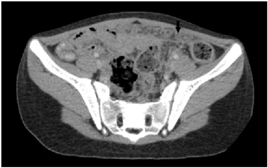
Results: The study included 28 males and 22 females, with a mean age of 8.2 ± 3.5 years. The most common clinical features were ascites (72%), abdominal pain (62%), and weight loss (40%). Diagnostic classification revealed 15 microbiologically confirmed, 25 histopathology-confirmed, and 10 probable cases. Ascites was the most frequent imaging finding on both ultrasound and CT. Other common findings included mesenteric lymphadenopathy, bowel wall thickening, and omental involvement. Stratified analysis demonstrated that ascites was prevalent across all diagnostic categories (80%, 72%, and 60%, respectively). Necrotic lymphadenopathy, bowel wall thickening, and omental thickening showed overlapping distributions among groups, with no statistically significant differences (p > 0.05). GeneXpert positivity was significantly associated with necrotic lymphadenopathy (p < 0.05).
Conclusion: Pediatric abdominal tuberculosis demonstrates a spectrum of overlapping imaging findings across diagnostic categories. While features such as ascites and necrotic lymphadenopathy are common, they are not specific to confirmed disease. Imaging plays an important supportive role, particularly in probable cases lacking microbiological confirmation. An integrated approach combining clinical, laboratory, and radiological findings is essential for timely diagnosis and management.
References
1. Pekcan S, Tana Aslan A, Kiper N, et al. Multicentric analysis of childhood tuberculosis in Turkey. Turk J Pediatr 2013; 55:121-9
2. Somu N, Vijayasekaran D, Ravikumar T, et al. Tuberculous disease in a pediatric referral centre: 16 years’ experience. Indian Pediatr. 1994; 31:1245–1249.
3. Delisle M, Seguin J, Zeilinski D, et al. Paediatric abdominal tuberculosis in developed countries: case series and literature review [published correction appears in Arch Dis Child. 2016 Apr;101(4):411]. Arch Dis Child. 2016; 101:253–258
4. Kapoor VK: Abdominal tuberculosis. Postgrad Med J. 1998, 74:459-67. 10.1136/pgmj.74.874.459
5. Handa U, Mundi I, Mohan S: Nodal tuberculosis revisited: a review . J Infect Dev Ctries. 2012, 6:6-12. 10.3855/jidc.2090
6. Rana S, Farooqui MR, Rana S, Anees A, Ahmad Z, Jairajpuri ZS: The role of laboratory investigations in evaluating abdominal tuberculosis. J Family Community Med. 2015, 22:152-7. 10.4103/2230-8229.163029
7. Sathar MA, Simjee AE, Coovadia YM, Soni PN, Moola SA, Insam B, Makumbi F: Ascitic fluid gamma interferon concentrations and adenosine deaminase activity in tuberculous peritonitis. Gut. 1995, 36:419- 21. 10.1136/gut.36.3.419
8. Nishal N, Arjun P, Arjun R, Ameer KA, Nair S, Mohan A: Diagnostic yield of CBNAAT in the diagnosis of extrapulmonary tuberculosis: A prospective observational study. Lung India. 2022, 39:443-8. 10.4103/lungindia.lungindia_165_22
9. Debi U, Ravisankar V, Prasad KK, Sinha SK, Sharma AK. Abdominal tuberculosis of the gastrointestinal tract: Revisited. World J Gastroenterol. 2014;20:14831–14840.
10. Sharma MP, Bhatia V. Abdominal tuberculosis. Indian J Med Res. 2004;120:305–315.
11. Rasheed S, Zinicola R, Watson D, Bajwa A, McDonald PJ. Intra-abdominal tuberculosis: A retrospective review of 218 cases. QJM. 2007;100:1–7.
12. Jain R, Sawhney S, Bhargava DK, Berry M. Diagnosis of abdominal tuberculosis: Sonographic findings in patients with early disease. AJR Am J Roentgenol. 1995;165:1391–1395.
13. Kedar RP, Shah PP, Shiralkar SR, Malde HM. Sonographic findings in gastrointestinal and peritoneal tuberculosis. Clin Radiol. 1994;49:24–29.
14. Burrill J, Williams CJ, Bain G, Conder G, Hine AL, Misra RR. Tuberculosis: A radiologic review. Radiographics. 2007;27:1255–1273.
15. Balthazar EJ, Gordon R, Hulnick D. Ileocecal tuberculosis: CT and radiologic evaluation. AJR Am J Roentgenol. 1990;154:499–503.
16. Horvath KD, Whelan RL. Intestinal tuberculosis: Return of an old disease. Am J Gastroenterol. 1998;93:692–696.
17. World Health Organization. Guidance for National Tuberculosis Programmes on the Management of Tuberculosis in Children. WHO; 2014.
Downloads
Published
Data Availability Statement
Available from the corresponding author upon reasonable request.
Issue
Section
License
Copyright (c) 2026 Dr. Sumaira Maqbool, Dr. Akib Arfee, Dr Syed Ruzina Firdose, Dr Mushtaq Wani

This work is licensed under a Creative Commons Attribution 4.0 International License.
This license requires that reusers give credit to the creator. It allows reusers to distribute, remix, adapt, and build upon the material in any medium or format, even for commercial purposes.


