Vol. 27 No. 1 (2026): Vol. 27 No. 1 (2026)
Russian Federation - Human and Artificial Intelligence in Radiology: Current Status, Evidence, Regulation, and Future Perspectives
https://doi.org/10.59667/sjoranm.v27i1.14
Zainab Magomedova1,2, Ekaterina S. Pershina1,2, Keivan Daneshvar3, Gerd Nöldge3, Frank Mosler3
Artificial intelligence (AI) has rapidly evolved into a transformative force in radiology, complementing human intelligence across the entire imaging workflow. Current applications range from image acquisition and reconstruction to automated detection, quantification, triage, and clinical decision support. Evidence to date demonstrates that AI systems can match or exceed human performance in narrowly defined tasks, particularly in pattern recognition and workflow optimization. However, robust prospective validation, demonstration of clinical impact, and proof of generalizability across institutions and populations remain limited.
----------------------------------------------------------------------------------------------
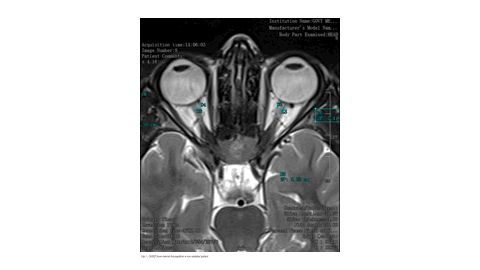
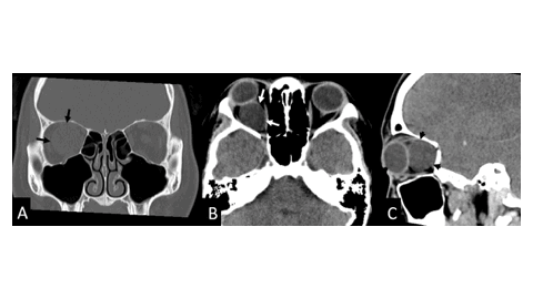
Chile - Optic Nerve Sheath Meningocele: a case report and review of the literature
https://doi.org/10.59667/sjoranm.v27i1.16
Ilson Sepúlveda Aguilar1*, Francisco Rivas-Rodriguez2
Optic nerve sheath meningocele (ONSM) is a rare condition with only a few cases reported in the medical literature. The etiology is unknown. The condition is characterized by an expansion of the cerebrospinal fluid space surrounding the optic nerve, without associated inflammation or the presence of orbital or cerebral neoplasms at the apex of the orbit. The condition is characterized by the absence of specific symptoms, with the most common being blurred vision and retro-orbital pain. We present the case of a young patient who was admitted to the emergency department at an external hospital. A clinical examination revealed painless right exophthalmos. No additional neurological symptoms were observed. A Computed Tomography (CT) scan and Magnetic Resonance Imaging (MRI) revealed an ONSM.
----------------------------------------------------------------------------------------------
https://doi.org/10.59667/sjoranm.v27i1.18
Onwuzu, Sobechukwu W I1; Uche-Nwankwo, Adanna Munachimsoaga1*; Ozoamalu, Chinemerem Sebastian1; Ozohili, Chikaosolu Vanessa1; Peter-Adim, Ezinne Stacey1; Solomon, Prisca Chinecherem1; Sopuluchukwu, Precious Ogechi1; Onwunta, Ikechukwu Emmanuel1
1Department of Medical Radiography and Radiological Sciences, University of Nigeria
Nigerian radiographers demonstrate moderate familiarity with AI but limited understanding of theranostics. While job security concerns persist, willingness to upskill is strong. Structured training, mentorship, and curriculum reform are essential to support professional adaptation and safeguard radiographers’ relevance in the era of AI and theranostics.
----------------------------------------------------------------------------------------------
India - Visual Journey through Tuberous Sclerosis Complex: Multisystem Imaging Insights
https://doi.org/10.59667/sjoranm.v27i1.20
Thiagarajan Veerappan1, Geetha Ganesan1, Kalpana Sivalingam1
1Barnard Institute of Radiology, Madras Medical College, Chennai, Tamil Nadu, India
Tuberous sclerosis complex (TSC) is a rare genetic disorder affecting several systems, characterized by hamartomatous lesions in the brain, kidneys, lungs, skin, and bones. Imaging plays a pivotal role in diagnosis and management. We report a case series of four patients exhibiting diverse clinical manifestations who received multimodal imaging from 2022 to 2024. The imaging findings were aligned with clinical and diagnostic criteria established by the 2012 International TSC Consensus guidelines. The cases had distinctive radiological characteristics of TSC, encompassing subependymal nodules, subependymal giant cell astrocytomas (SEGAs), renal angiomyolipomas, pulmonary lymphangioleiomyomatosis (LAM), cutaneous lesions, and skeletal anomalies. Cross-sectional imaging facilitated precise diagnosis and directed therapies, including embolization, for renal pseudoaneurysms. The series highlights the significance of a thorough imaging strategy in recognizing both typical and incidental characteristics of TSC. Prompt identification enables swift diagnosis, focused treatment, and long term surveillance.
----------------------------------------------------------------------------------------------
https://doi.org/10.59667/sjoranm.v27i1.22
Omair Ashraf Shah1, Mudhabir Ashraf2, Akib Arfee1*, Sumaira Maqbool3
1Department of Radiodiagnosis and Imaging, Govt. Medical College Srinagar, India
2Department of Anaesthesia and Critical Care, Govt. Medical College Srinagar, India
Sedation significantly influences ONSD measurements on MRI in pediatric patients. These findings highlight the need for careful consideration of sedation status when using ONSD as a surrogate for ICP. Age-related trends further highlight the importance of using adjusted reference values.
----------------------------------------------------------------------------------------------
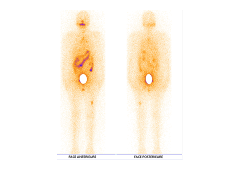
https://doi.org/10.59667/sjoranm.v27i1.24
Halima Batani1*, Hafsa Bensimimou1, Abdel Amide Gbadamassi1,2, Zakaria Ouassafrar1, Amal Guensi1
1Nuclear Medicine Department, Ibn Rochd University Hospital Center, Casablanca, Morocco
[18F]FDG PET/CT is an essential tool in the diagnostic and therapeutic evaluation of poorly differentiated follicular-origin thyroid carcinomas. Its contribution is pivotal in guiding clinical decision-making. Moreover, surgical management of isolated bone metastases can offer a genuine opportunity for durable disease control.
----------------------------------------------------------------------------------------------
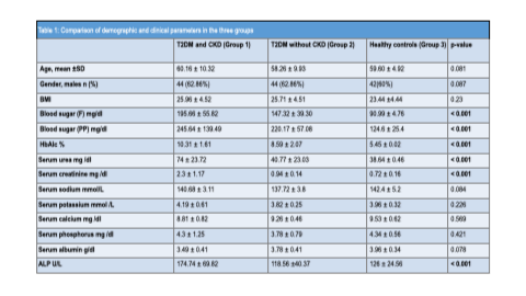
https://doi.org/10.59667/sjoranm.v27i1.26
Surjeet Chandail1, Arshed Hussain Parry1*, Majid Jehangir1, Syeed Aalishan Fatima1, Seema Qayoom2
Patients with T2DM, particularly those with concomitant CKD, exhibit a substantially higher prevalence of osteopenia and osteoporosis compared to non-diabetic individuals. These findings emphasize the need for routine bone health assessment and early osteoporosis screening in diabetic patients to reduce fracture risk and improve clinical outcomes.
----------------------------------------------------------------------------------------------
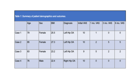
USA - Multivessel Transcatheter Arterial Embolization to Treat Hip Osteoarthritis: A Pilot Case Series
https://doi.org/10.59667/sjoranm.v27i1.28
Prem Krishnamurthy1*, Nishanth Konduru1, Siddhartha Rao1
1Rao Clinic, Cary, North Carolina, United States
Chronic hip pain due to osteoarthritis (OA) is a prevalent source of disability in older adults. While total hip arthroplasty (THA) remains the standard treatment for end-stage disease, many patients are not surgical candidates due to comorbidities or personal preferences. Transcatheter Arterial Embolization (TAE) of the hip has emerged as a minimally invasive treatment for OA-related pain, but further evidence is warranted to establish its role in treatment pathways.
----------------------------------------------------------------------------------------------