
The Multiple Myeloma
Extramedullary Manifestations
DOI:
https://doi.org/10.59667/sjoranm.v6i1.14Keywords:
multiple myeloma, extramedullary manifestations, testicular blood barrierAbstract
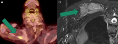
The second most common hematologic malignancy is the clonal proliferation of neoplastic plasma cells within the bone marrow. There is the presence of monoclonal immunoglobulins in the serum and/or urine. This results in anemia, myelosuppression, bone destruction, and clinical consequences of para-proteinemia on kidney function and other organ systems. The disease manifests through the acronym CRAB (hypercalcemia, renal impairment, anemia, and bone lesions). Less frequent manifestations of multiple myeloma are of extramedullary localizations. Myeloma cells can become independent of the bone marrow microenvironment, circulate freely in the blood, and infiltrate organs. This results in a high-risk state characterized by increased proliferation, evasion of apoptosis, and treatment resistance. It can affect any area of tissue. Most commonly it involves the pleura, lymph nodes, chest wall, liver, skin/soft tissue, lungs, CNS, genitourinary system, breast and pancreas. In patients with confirmed multiple myeloma, the diagnosis of extramedullary involvement is typically established by the presence of pathological soft tissue masses using radiological methods such as computed tomography (CT) scan, positron emission tomography/CT (PET/CT), magnetic resonance imaging (MRI), or ultrasound, along with biopsy or physical examination. The molecular mechanisms underlying the development of extramedullary multiple myeloma (EMM) have not been fully defined. Various cytogenetic abnormalities are observed, and some studies have generated genomic sequencing profiles that distinguish EMM from classic multiple myeloma. While plasma cell leukemia (PCL) and central nervous system (CNS) EMM indicate a poor prognosis, outcomes for other manifestations can be highly heterogeneous. Sensitive imaging modalities including PET/CT and MRI (Fig.1) are integral components of diagnosis and response assessment. Patients with extramedullary multiple myeloma (EMM) have a clear survival disadvantage.
References
Catto MB, Safranauskas R, Datoguia TS, et al.
Cytogenetic findings in testicular relapse of
multiple myeloma: case report and literature
review. Hematol Transfus Cell Ther 2024. DOI:
0 . 1 0 1 6 / j . h t c t . 2 0 2 3 . 1 2 . 0 0 2 . h t t p s : / /
www.ncbi.nlm.nih.gov/pubmed/38402033 https://
www.sciencedirect.com/science/article/pii/
S2531137924000063?via%3Dihub
Strauss D, Sachpekidis C, Dapunt U, Gold
schmidt H, Dimitrakopoulou-Strauss A. Unusual
Extramedullary Manifestation in Multiple
Myeloma: Bilateral Synchronous Testicular Infiltra-
tion. Clin Nucl Med 2023;48(2):e76-e77. DOI:
1097/RLU.0000000000004496. https://
www.ncbi.nlm.nih.gov/pubmed/36399719
Doshi K, Abedrabo S, Bitran J, Asado N. A Case
of Recurrent Multiple Myeloma as Testicular
Plasmacytoma Without Systemic Disease. Cureus
;15(6):e40785. DOI: 10.7759/cureus.40785.
https://www.ncbi.nlm.nih.gov/pubmed/37485101
https://www.ncbi.nlm.nih.gov/pmc/articles/
P M C 1 0 3 6 2 5 2 7 / p d f /
cureus-0015-00000040785.pdf
Mohan M, Yarlagadda N, Szabo A, Singh A, Pina
Oviedo S, Schinke C. Clinical characteristics of
testicular extramedullary involvement in multiple
myeloma. Am J Hematol 2021;96(3):E77-E81.
D O I : 1 0 . 1 0 0 2 / a j h . 2 6 0 7 2 . h t t p s : / /
www.ncbi.nlm.nih.gov/pubmed/33338289
Bhutani M, Foureau DM, Atrash S, Voorhees PM,
Usmani SZ. Extramedullary multiple myeloma.
L e u k e m i a 2 0 2 0 ; 3 4 ( 1 ) : 1 - 2 0 . ( h t t p s : / /
www.nature.com/articles/s41375-019-0660-0).
Yamashita K, Horiuchi T, Hayashida A, Tachibana
H, Toki D, Kondo T. Multiple myeloma with
testicular involvement: a case report. Urology
Case Reports 2019;26:100971. (https://
w w w. n c b i . n l m . n i h . g o v / p m c / a r t i c l e s /
PMC6659131/pdf/main.pdf).
Sevcikova S, Minarik J, Stork M, Jelinek T, Pour L,
Hajek R. Extramedullary disease in multiple
myeloma–controversies and future directions.
Blood reviews 2019;36:32-39. (https://
www.sciencedirect.com/science/article/pii/
S0268960X18300961?via%3Dihub).
Ormond Filho AG, Carneiro BC, Pastore D, et al.
Whole-body imaging of multiple myeloma:
diagnostic criteria. Radiographics 2019;39(4):
-1097. (https://pubs.rsna.org/doi/10.1148/
rg . 2 0 1 9 1 8 0 0 9 6 ? u r l _ v e r = Z 3 9 . 8 8 - 2 0 0 3 -
&rfr_id=ori:rid:crossref.org&rfr_dat=cr_pub%20%
pubmed).
Rajkumar SV. Updated diagnostic criteria and
staging system for multiple myeloma. American
Society of Clinical Oncology Educational Book
;36:e418-e423.
Pasmantier MW, Azar HA. Extraskeletal spread in
multiple plasma cell myeloma: A review of 57
autopsied cases. Cancer 1969;23(1):167-174.
Downloads
Published
Issue
Section
License
Copyright (c) 2024 Keivan Daneshvar, Wolfram Andreas Bosbach, Nando Mertineit, Gerd Nöldge; Frank Mosler

This work is licensed under a Creative Commons Attribution 4.0 International License.
This license requires that reusers give credit to the creator. It allows reusers to distribute, remix, adapt, and build upon the material in any medium or format, even for commercial purposes.


