Vol. 4 No. 1 (2024): Vol. 04 No. 1 (2024)

Purpose:
To compare the efficacy of computed tomography (CT) and magnetic resonance imaging (MRI) in evaluating cystic renal masses using the Bosniak classification system.
Materials and Methods:
Between January 2010 and December 2020, a total of 844 patients with suspected renal masses underwent total or partial nephrectomy at the Department of Urology, Heidelberg University Clinic. Among them, 123 patients presented with cystic renal masses. Out of this cohort, 15 patients underwent preoperative examinations using both MRI and CT within a 6-week timeframe. These examinations were retrospectively analyzed by two radiologists employing the Bosniak classification system. Each lesion was assessed based on CT images for lesion localization, volume, number, and thickness of septa/wall, calcification, presence of enhancing soft-tissue components, and lesion density. Pathologic correlation was available for all lesions.
Results:
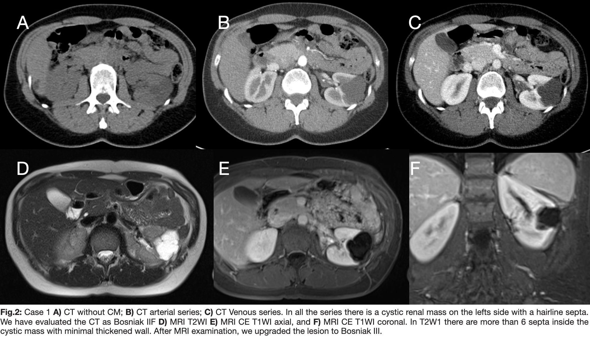
Out of the 844 patients, 15 underwent both CT and MR imaging examinations (male-to-female ratio: 2.75, age range: 43-75 years; mean age: 60.5 years). Based on CT images, there were one category I, one category II, three category IIF, three category III, and seven category IV lesions. Findings based on CT and MR images were concordant in eleven (73.3%) renal lesions, while in 4 (26.7%) masses, differences were observed. In three (20%) cases, MR imaging revealed more septa/wall than CT, resulting in an upgrade of the classification at MR imaging. In one (6.7%) lesion, MR imaging showed no wall and/or septa thickness compared with CT, leading to a classification downgrade. MR imaging results led to a cyst classification upgrade in three masses (from category II to III, n=1; IIF to III, n=2) and downgrade in one lesion (from category IIF to II). Pathologically, 11 lesions (73.3%) were malignant (9 Cystic Clear cell carcinoma and 2 papillary renal cell carcinoma), while 4 lesions were renal cysts without malignancy.
Conclusion:
The majority of findings from both CT and MR imaging were similar in suspected cystic renal masses. MR imaging demonstrated additional septa/wall or septa/wall thickening, potentially upgrading lesions from borderline category IIF to III compared to CT. This distinction may contribute to improved case management.
----------------------------------------
The Transarterial Embolization of Visceral Artery AneurysmsThe management of an aneurysm or pseudoaneurysm, including decisions regarding how, when, and with what method it should be treated, must always be determined through interdisciplinary consensus grounded in a thorough understanding of the existing literature. Consequently, today's preferred approach is embolization therapy or covered stenting. This method, performed in a minimally invasive manner, has been well-established due to its methodical low invasiveness and high effectiveness in achieving both technical and clinical success. It stands as a prominent choice among the available surgical and minimally invasive therapy options. The procedure, executed with advanced technology, utilizes the latest catheters and wires, coupled with expertise in various embolization methods. This comprehensive approach enables the treatment of acute bleeding in emergency cases, ensuring a nuanced and effective response.