Osteoid Osteoma
Synopsis of clinical, radiological, and therapeutical relevance of this rare entity
DOI:
https://doi.org/10.59667/sjoranm.v.1i.15Keywords:
Osteoid osteoma, radio frequency ablation, stereotactic navigation with CT-guidance, benign osseous tumor, excochleation, imaging of nidusAbstract
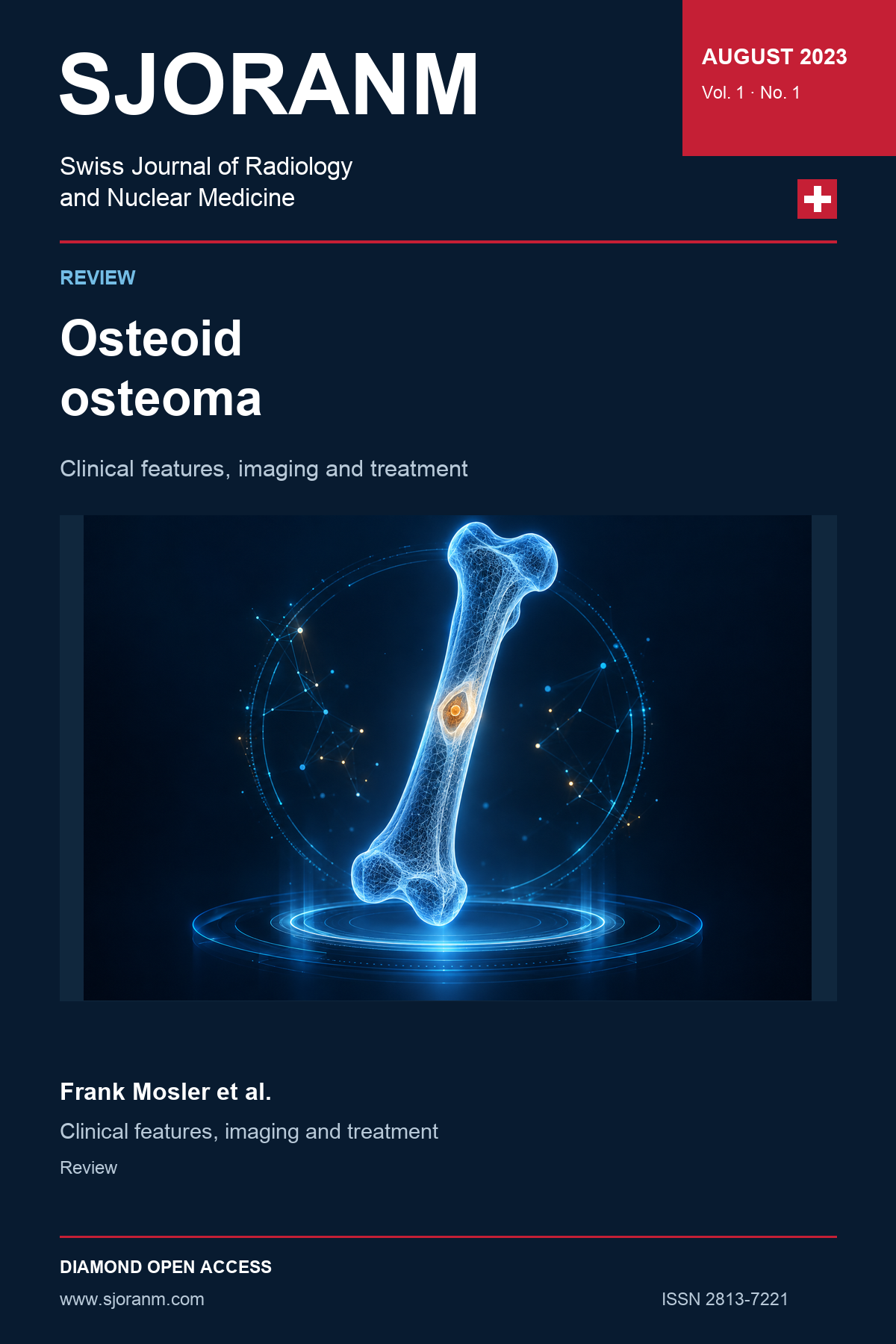
Characteristic of the osteoid osteoma is the so-called nidus enclosed in the tumor and produces a typical picture on X-ray. Since most physicians have little experience with the clinical picture of osteoid osteoma, this is an important reason for the often-long anamnesis time for osteoid osteoma (OO). The period of formation of OO includes the phase of strongest bone growth in childhood and adolescence. The most characteristic clinical symptom is nocturnal attacks of pain around the tumor, which occur independently of preceding physical activity and respond strikingly well to non-steroidal anti-inflammatory drugs (aspirin test). OO most frequently affects the long tubular bones of the lower extremities. Fifty percent of OO are found in the femur and tibia preferentially occurring in the corticalis of metaphysis and diaphysis. This is followed by the long tubular bones of the upper extremity and the short tubular bones. However, any other bone can also be affected. Differentially, osteoblastoma or osteosarcoma must be considered first and foremost. Most widely accepted therapy options are open surgery with en-bloc resection of the tumor, excochleation,minimally invasive percutaneous CT-guided radio or laser ablation. A conservative management by pharmaceutical pain therapy should be reserved for cases in which surgery must be refused either because of the precarious position of the nidus or because the patient's general condition does not permit it, or the patient does not consent to surgical interventions.References
1. JAFFE HL. "OSTEOID-OSTEOMA" A BENIGN OSTEOBLASTIC TUMOR COMPOSED OF OSTEOID AND ATYPICAL BONE. Arch Surg 1935;31(5):709-728. (Original article) (In English). DOI: https://doi.org/10.1001/archsurg.1935.01180170034003. DOI: https://doi.org/10.1001/archsurg.1935.01180170034003
2. Dahlin DC. Giant-cell-bearing lesions of bone of the hands. Hand Clin 1987;3(2):291-7. (https://www.ncbi.nlm.nih.gov/pubmed/3584256). DOI: https://doi.org/10.1016/S0749-0712(21)00659-4
3. Schajowicz F. Cartilage-forming tumors. Tumors and tumorlike lesions of bone: pathology, radiology, and treatment 1994:141-256. DOI: https://doi.org/10.1007/978-3-642-49954-8_3
4. Voggenreiter G, Klaes W, Nast-Kolb D. Das Osteoidosteom ein diagnostisches und therapeutisches Problem? Der Chirurg 2000;3(71):319-325.
5. Freyschmidt J, Ostertag H, Jundt G. Bone tumors. Clinical experience-radiology-pathology; 2. rev. and enl. ed.; Knochentumoren. Klinik-Radiologie-Pathologie. 1998.
6. Campanacci M, Ruggieri P, Gasbarrini A, Ferraro A, Campanacci L. Osteoid osteoma. Direct visual identification and intralesional excision of the nidus with minimal removal of bone. J Bone Joint Surg Br 1999;81(5):814-20. DOI: https://doi.org/10.1302/0301-620x.81b5.9313. DOI: https://doi.org/10.1302/0301-620X.81B5.0810814
7. Kayser M, Muhr G. Eighteen-year anamnesis of osteoid osteoma--a diagnostic problem? Arch Orthop Trauma Surg (1978) 1988;107(1):27-30. DOI: https://doi.org/10.1007/BF00463521. DOI: https://doi.org/10.1007/BF00463521
8. Adler C-P. Knochenkrankheiten: Diagnostik makroskopischer, histologischer und radiologischer Strukturveränderungen des Skeletts: Springer-Verlag, 2013.
9. Heuck A, Stabler A, Wortler K, Steinborn M. [Benign bone-forming tumors]. Radiologe 2001;41(7):540-7. DOI: https://doi.org/10.1007/s001170170144. DOI: https://doi.org/10.1007/s001170170144
10. Wespe R, Aebi M. [Osteoid osteoma of the spine and pelvic area]. Schweiz Med Wochenschr 1990;120(30):1102-8. (https://www.ncbi.nlm.nih.gov/pubmed/2392661).
11. Schulman L, Dorfman HD. Nerve fibers in osteoid osteoma. J Bone Joint Surg Am 1970;52(7):1351-6. (https://www.ncbi.nlm.nih.gov/pubmed/4097043). DOI: https://doi.org/10.2106/00004623-197052070-00008
12. Sherman M, McFarland Jr G. Mechanism of pain in osteoid osteomas. Southern medical journal 1965;58:163-166. DOI: https://doi.org/10.1097/00007611-196502000-00005
13. Esquerdo J, Fernandez C, Gomar F. Pain in osteoid osteoma: histological facts. Acta Orthopaedica Scandinavica 1976;47(5):520-524. DOI: https://doi.org/10.3109/17453677608988731
14. Berning W, Freyschmidt J, Wiens J. Zur perkutanen therapie des osteoidosteoms. Der Unfallchirurg 1997;100:536-540. (https://link.springer.com/article/10.1007/s001130050154). DOI: https://doi.org/10.1007/s001130050154
15. Resnick D, Niwayama G. Diagnosis of bone and joint disorders. 1987.
16. Leonhardt J, Bastian L, Rosenthal H, Laenger F, Wippermann B. Post-traumatic osteoid osteoma. Case report and review of the literature. Der Unfallchirurg 2001;104(6):553-556. (https://link.springer.com/article/10.1007/s001130170120). DOI: https://doi.org/10.1007/s001130170120
17. Assenmacher S, Voggenreiter G, Klaes W, Nast-Kolb D. [Osteoid osteoma--a diagnostic and therapeutic problem?]. Chirurg 2000;71(3):319-25. DOI: https://doi.org/10.1007/s001040051057. DOI: https://doi.org/10.1007/s001040051057
18. Allen S, Saifuddin A. Imaging of intra-articular osteoid osteoma. Clinical radiology 2003;58(11):845-852. (https://www.clinicalradiologyonline.net/article/S0009-9260(03)00213-7/fulltext). DOI: https://doi.org/10.1016/S0009-9260(03)00213-7
19. Gamba JL, Martinez S, Apple J, Harrelson JM, Nunley JA. Computed tomography of axial skeletal osteoid osteomas. AJR Am J Roentgenol 1984;142(4):769-72. DOI: https://doi.org/10.2214/ajr.142.4.769. DOI: https://doi.org/10.2214/ajr.142.4.769
20. Frassica FJ, Waltrip RL, Sponseller PD, Ma LD, McCarthy Jr EF. Clinicopathologic features and treatment of osteoid osteoma and osteoblastoma in children and adolescents. Orthopedic Clinics of North America 1996;27(3):559-574. DOI: https://doi.org/10.1016/S0030-5898(20)32101-5
21. Yildirim G, Karakas HM, Yilmaz B. Uncooled microwave ablation of osteoid osteoma: New approaches to an old problem. North Clin Istanb 2022;9(5):524-529. DOI: https://doi.org/10.14744/nci.2021.26675. DOI: https://doi.org/10.14744/nci.2021.26675
22. Helms CA, Hattner RS, Vogler JB, 3rd. Osteoid osteoma: radionuclide diagnosis. Radiology 1984;151(3):779-84. DOI: https://doi.org/10.1148/radiology.151.3.6232642. DOI: https://doi.org/10.1148/radiology.151.3.6232642
23. ZHOU X, HUANG T-t, YE Y. MRI Diagnosis of Osteoid Osteoma in Joint Capsule. CHINESE JOURNAL OF CT AND MRI 2022;20(09):155.
24. Assoun J, Richardi G, Railhac JJ, et al. Osteoid osteoma: MR imaging versus CT. Radiology 1994;191(1):217-23. DOI: https://doi.org/10.1148/radiology.191.1.8134575. DOI: https://doi.org/10.1148/radiology.191.1.8134575
25. Lee DH, Malawer MM. Staging and treatment of primary and persistent (recurrent) osteoid osteoma. Evaluation of intraoperative nuclear scanning, tetracycline fluorescence, and tomography. Clinical orthopaedics and related research 1992(281):229-238. DOI: https://doi.org/10.1097/00003086-199208000-00039
26. Buhler M, Binkert C, Exner GU. Osteoid osteoma: technique of computed tomography-controlled percutaneous resection using standard equipment available in most orthopaedic operating rooms. Arch Orthop Trauma Surg 2001;121(8):458-61. DOI: https://doi.org/10.1007/s004020100264. DOI: https://doi.org/10.1007/s004020100264
27. Adam G, Keulers P, Vorwerk D, Heller K-D, Füzesi L, Günther R. Perkutane CT-gesteuerte Behandlung von Osteoidosteomen: kombiniertes Vorgehen mit einem Hohlbohrer und nachfolgender Äthanolinjektion. RöFo-Fortschritte auf dem Gebiet der Röntgenstrahlen und der bildgebenden Verfahren: © Georg Thieme Verlag Stuttgart· New York; 1995:232-235. DOI: https://doi.org/10.1055/s-2007-1015871
28. Gangi A, Dietemann JL, Guth S, et al. Percutaneous laser photocoagulation of spinal osteoid osteomas under CT guidance. AJNR Am J Neuroradiol 1998;19(10):1955-8. (https://www.ncbi.nlm.nih.gov/pubmed/9874556).
29. Wasserlauf B, Gossett J, Rosenthal DI, Levine WN. Osteoid osteoma of the glenoid: minimally invasive treatment. American Journal of Orthopedics (Belle Mead, NJ) 2003;32(8):405-407.
30. Donahue F, Ahmad A, Mnaymneh W, Pevsner NH. Osteoid osteoma. Computed tomography guided percutaneous excision. Clin Orthop Relat Res 1999(366):191-6. (https://www.ncbi.nlm.nih.gov/pubmed/10627735). DOI: https://doi.org/10.1097/00003086-199909000-00025
31. Sans N, Galy-Fourcade D, Assoun J, et al. Osteoid osteoma: CT-guided percutaneous resection and follow-up in 38 patients. Radiology 1999;212(3):687-92. DOI: https://doi.org/10.1148/radiology.212.3.r99se06687. DOI: https://doi.org/10.1148/radiology.212.3.r99se06687
32. Rosenthal DI, Springfield DS, Gebhardt MC, Rosenberg AE, Mankin HJ. Osteoid osteoma: percutaneous radio-frequency ablation. Radiology 1995;197(2):451-454. DOI: https://doi.org/10.1148/radiology.197.2.7480692
33. Cantwell CP, Obyrne J, Eustace S. Current trends in treatment of osteoid osteoma with an emphasis on radiofrequency ablation. European radiology 2004;14:607-617. DOI: https://doi.org/10.1007/s00330-003-2171-6
34. Kneisl JS, Simon MA. Medical management compared with operative treatment for osteoid-osteoma. J Bone Joint Surg Am 1992;74(2):179-85. (https://www.ncbi.nlm.nih.gov/pubmed/1541612). DOI: https://doi.org/10.2106/00004623-199274020-00004
35. Ilyas I, Younge DA. Medical management of osteoid osteoma. Can J Surg 2002;45(6):435-7. (https://www.ncbi.nlm.nih.gov/pubmed/12500919).
36. Yalcinkaya U, Doganavsargil B, Sezak M, et al. Clinical and morphological characteristics of osteoid osteoma and osteoblastoma: a retrospective single-center analysis of 204 patients. Ann Diagn Pathol 2014;18(6):319-325. DOI: https://doi.org/10.1016/j.anndiagpath.2014.08.006. DOI: https://doi.org/10.1016/j.anndiagpath.2014.08.006
37. Fittall MW, Mifsud W, Pillay N, et al. Recurrent rearrangements of FOS and FOSB define osteoblastoma. Nat Commun 2018;9:2150. DOI: https://doi.org/10.1038/s41467-018-04530-z. DOI: https://doi.org/10.1038/s41467-018-04530-z
38. Akhlaghpoor S, Aziz Ahari A, Ahmadi SA, et al. Histological evaluation of drill fragments obtained during osteoid osteoma radiofrequency ablation. Skeletal Radiol 2010;39(5):451-455. DOI: https://doi.org/10.1007/s00256-010-0901-9. DOI: https://doi.org/10.1007/s00256-010-0901-9
Downloads
Published
Data Availability Statement
Not applicable
Issue
Section
License
Copyright (c) 2023 Frank Mosler

This work is licensed under a Creative Commons Attribution 4.0 International License.
This license requires that reusers give credit to the creator. It allows reusers to distribute, remix, adapt, and build upon the material in any medium or format, even for commercial purposes.
 LinkedIn ↗
 X ↗
 VK ↗
 Truth Social ↗
 Bluesky ↗
 YouTube ↗