Vol. 30 No. 1 (2026): Vol. 30 No. 1 (2026)

The Evaluation of focal breast lesions using ultrasound elastography with FNAC and/or histopathology correlation among patients visiting breast ultrasound and mammography units at Tikur Anbessa Specialized Teaching Hospital, Ethiopia, August, 2025
Background: Breast cancer continues to be a major cause of illness and death worldwide, including in Ethiopia. Traditionally, diagnosis in Ethiopia has relied on clinical palpation, mammography, and B-mode ultrasonography (US). Histopathological examination continues to be the gold standard for definitive diagnosis. Recently, elastography has emerged as a promising adjunct to B-mode ultrasonography, enhancing specificity and aiding in the early detection of breast cancer.
Methodology: A prospective analytical cross-sectional study was carried out at Tikur Anbessa Specialized Hospital (TASH) between December 1, 2024 and April 30, 2025.
Result: The study involved 100 patients, 72% had malignant breast lesions, 26% benign, and 2% atypical, with invasive carcinoma and fibroadenoma being the most common. BI-RADS alone showed high sensitivity (98.6%) but low specificity (7.7%). Adding elasticity scoring and strain ratio improved diagnostic accuracy. Elasticity scoring achieved 96% sensitivity and 69% specificity, while strain ratio had 98.6% sensitivity and 54% specificity. Serial testing method provided a more balanced approach with 93% sensitivity, 86.9% specificity, and 93.5% accuracy, reducing false positives while maintaining diagnostic strength.
Conclusion: This study highlights the clinical value of integrating BI-RADS, elasticity scoring, and strain ratio in the ultrasound assessment of breast lesions.
Keywords: BIRADS 3, BIRADS 4, breast lesion, cytology, elasticity scoring, elastography, histopathology, sensitivity and specificity, strain ratio, ultrasound
----------------------------------------
Contribution of [18F]FDG PET/CT in the detection of a penile metastasis from pulmonary squamous cell carcinoma
Introduction: Lung cancer remains one of the leading causes of cancer-related mortality worldwide and is characterized by marked biological aggressiveness, resulting in frequent and often early metastases. Positron emission tomography combined with computed tomography (PET/CT) using ¹⁸F-fluorodeoxyglucose ([18F]FDG) has become an essential tool in the staging workup of non–small cell lung cancers, particularly squamous cell carcinomas, which are known for their high glucose avidity. While common metastatic sites include lymph nodes, bone, liver, brain, and adrenal glands, penile metastases are exceptionally rare and are associated with a poor prognosis.
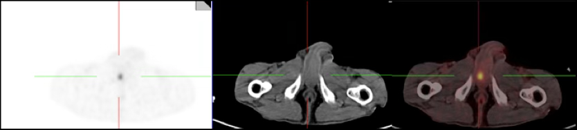
Case report: We report the case of a 65-year-old patient with a long history of heavy smoking, who presented with a persistent cough associated with chest pain. Thoracic imaging revealed a mass in the left lower lobe, and histological and immunohistochemical analyses confirmed the diagnosis of pulmonary squamous cell carcinoma. As part of the initial staging workup, an ¹⁸F-FDG PET/CT scan was performed. This examination demonstrated an intensely hypermetabolic primary lung lesion with extensive mediastinal involvement, associated with adrenal and bone metastases. Incidentally and in the absence of any related symptoms, a well-circumscribed hypermetabolic focus was detected within the corpus cavernosum, consistent with a penile metastasis. This finding allowed the disease to be classified as stage IV and led to an adaptation of the therapeutic strategy.
Conclusion: This case highlights the major contribution of [18F]FDG PET/CT in the initial staging of lung cancers by enabling accurate disease staging and the detection of rare, clinically silent metastatic sites. Early identification of such metastases prevents the initiation of inappropriate curative-intent treatments and guides clinicians toward an appropriate, predominantly palliative therapeutic approach, thereby optimizing patient care.