The Contribution of Hybrid SPECT CT Imaging with [99mTc] Tc-MDP in Battered Woman Syndrome
A Case Study
DOI:
https://doi.org/10.59667/sjoranm.v23i1.20Keywords:
Battered woman syndrome, Bone scintigraphy, Hybrid SPECT/CT, TraumaAbstract
Introduction: Violence against women is a widespread health problem that knows no racial or social boundaries. It is generally difficult for forensic investigators to obtain medical evidence of violence, particularly in cases where the victims are assessed several months after the event. Bone scintigraphy can be used to identify and document trauma related to acts of torture, and it can also serve as valid evidence in court.
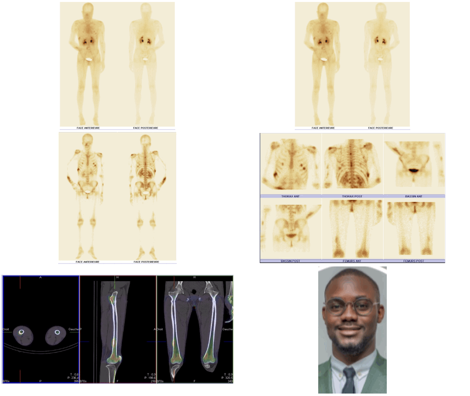
Case report: The bone scan revealed multiple areas of tissue uptake in the early phase, associated with bone uptake in the late phase involving both the axial and peripheral skeleton. In addition, several areas of bone uptake were observed in the late phase without corresponding tissue uptake in the early phase. The scintigraphic pattern demonstrated that the patient's bone lesions were of different ages. Based on these findings, early tissue uptake associated with late bone uptake was consistent with recent post-traumatic lesions, whereas late bone uptake without early tissue uptake was consistent with old post-traumatic lesions. The distribution of certain skeletal lesions suggested a defensive posture adopted by the victim.
Conclusion: In the context of a forensic investigation, bone scintigraphy is a valuable tool that not only detects bone lesions in patients suspected of physical assault, whether or not they present clinical signs, but also helps to determine when the injury occurred.
References
1. Ozkalipci O, Unuvar U, Sahin U, Irencin S, Fincanci SK. A significant diagnostic method in torture investigation: bone scintigraphy. Forensic Sci Int. 2013 Mar 10;226(1-3):142-5. https://doi.org/10.1016/j.forsciint.2012.12.019
2. Boeisa AN, Alghanim HA, Almutlaq A, Al-Saeed M, Alshaikhmubarak M. Occult Intertrochanteric Fracture Detected by Bone Scan Imaging: A Case Report. Cureus. 2024;18;16(7):e64815. doi: https://doi.org/10.7759/cureus.64815
3. Lok V, Tunca M, Kumanlioğlu K, Kapkin E, Dirik G. Bone scintigraphy as clue to previous torture. Lancet. 1991;337(8745):846-7. https://doi.org/10.1016/0140-6736(91)92548-g
4. Lok V, Tunca M, Kapkin E, et al. Bone scintigraphy as an evidence of previous torture: evidenced of 62 patients. In: Human Rights Foundation of Turkey (HRFT) Treatment and Rehabilitation Centers Report 1994. Ankara: HRFT Publications; 1995. pp. 91-96. Available from: https://en.tihv.org.tr/wp-content/uploads/2020/09/1994-treatment-centers-report.pdf
5. Mirzaei S, Knoll P, Lipp RW, Wenzel T, Koriska K, Köhn H. Bone scintigraphy in screening of torture survivors. Lancet. 1998;352(9132):949-51. https://doi.org/10.1016/s0140-6736(98)05049-1
6. Société Française de Médecine Nucléaire et Imagerie Moléculaire (SFMN). Guide pour la rédaction de protocole pour la scintigraphie osseuse. Version 2.0. Paris: SFMN; 2012. Available from: https://www.cnp-mn.fr/wp-content/uploads/2022/09/Os-V2.0.pdf
7. Blangis F, Poullaouec C, Launay E, Vabres N, Sadones F, Eugène T, Cohen JF, Chalumeau M, Gras-Le Guen C. Bone scintigraphy after a negative radiological skeletal survey improves the detection rate of inflicted skeletal injury in children. Front Pediatr. 2020;8:498. https://doi.org/doi:10.3389/fped.2020.00498
8. Spitz J, Becker C, Tittel K, Weigand H. Die klinische Relevanz der Ganzkörperskelettszintigraphie bei mehrfachverletzten und polytraumatisierten Patienten [Clinical relevance of whole body skeletal scintigraphy in multiple injury and polytrauma patients]. Unfallchirurgie. 1992 Jun;18(3):133-47. https://doi.org/10.1007/BF02588265
9. Heinrich SD, Gallagher D, Harris M, Nadell JM. Undiagnosed fractures in severely injured children and young adults. Identification with technetium imaging. J Bone Joint Surg Am. 1994 Apr;76(4):561-72. https://doi.org/10.2106/00004623-199404000-00011
10. Palestro CJ, Love C, Schneider R. The evolution of nuclear medicine and the musculoskeletal system. Radiol Clin North Am. 2009 May;47(3):505-32. https://doi.org/10.1016/j.rcl.2009.01.006
11. Offiah A, van Rijn RR, Perez-Rossello JM, Kleinman PK. Skeletal imaging of child abuse (non-accidental injury). Pediatr Radiol. 2009 May;39(5):461-70. Epub 2009 Feb 24. PMID: 19238374. https://doi.org/10.1007/s00247-009-1157-1
12. Holder LE, Schwarz C, Wernicke PG, Michael RH. Radionuclide bone imaging in the early detection of fractures of the proximal femur (hip): multifactorial analysis. Radiology. 1990 Feb;174(2):509-15. https://doi.org/10.1148/radiology.174.2.2404320
13. Lee E, Worsley DF. Role of radionuclide imaging in the orthopedic patient. Orthop Clin N Am. 2006;37:485–501. https://doi.org/10.1016/j.ocl.2006.04.003
14. Scheyerer MJ, Pietsch C, Zimmermann SM, Osterhoff G, Simmen HP, Werner CM. SPECT/CT for imaging of the spine and pelvis in clinical routine: a physician's perspective of the adoption of SPECT/CT in a clinical setting with a focus on trauma surgery. Eur J Nucl Med Mol Imaging. 2014;41 Suppl 1:S59-66. https://doi.org/10.1007/s00259-013-2554-0

Downloads
Published
Data Availability Statement
NA
License
Copyright (c) 2025 Abdel Amide Gbadamassi, Hafsa Bensimimoun, Halima Batani, Mohammed Amine Daerqaoui, Hicham Benyaich, Zakaria Ouassafrar, Amal Guensi

This work is licensed under a Creative Commons Attribution 4.0 International License.
This license requires that reusers give credit to the creator. It allows reusers to distribute, remix, adapt, and build upon the material in any medium or format, even for commercial purposes.


