Vol. 8 No. 1 (2024): Vol. 08 No. 1 (2024)
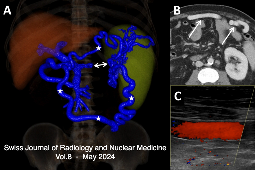
A 46-year-old man presented to the emergency department because of relapsing abdominal pain in the left abdominal quadrants. At admission, he was in good general condition and his vital signs were normal. The only abnormal laboratory findings were a C-reactive protein of 30 mg/L and thrombocytes of 127 G/L. An abdominal computed tomography (CT) scan showed a thrombosis of the distal splenic vein (panel A, double arrow) with the presence of a superior and an inferior spleno-mesenteric collateral (panel A, asterisks). The axial CT scan (panel V, arrows) and Doppler sonography (panel C) showed that the thick collaterals developed below the abdominal wall. A gastroscopy showed stage I fundic varices, but no esophageal varices. (1-10) This patient was diagnosed with a smoldering multiple myeloma 5 years earlier with regular follow-up in the hemato-oncology department. No other pro-thrombotic conditions were found and in a follow-up CT scan 6 months later the findings were unchanged despite anticoagulation. Splenic vein thrombosis is frequently diagnosed in the chronic stage, when collaterals have already developed (11-18). In this case, the presence of two major spontaneous shunts between the splenic vein and the superior mesenteric vein is preventing complications (19-23) such as porto-systemic encephalopathy and variceal bleeding.
-------------------------------------------------
The Role of 99mTc-HMDP Bone Scintigraphy in the Management of Neurogenic Para- Osteo-Arthropathies (NAOP)Neurogenic para-osteo-arthropathies (NAOP) are characterized by ectopic juxta-articular peri- or para-osseous ossifications. Diagnosis of these conditions is often delayed. They are frequently associated with central neurological disorders and are rarely observed in cases of peripheral neuropathy. Bone scintigraphy is crucial for determining the maturity of the ossification, optimizing surgical interventions, and preventing recurrence.
----------------------------------------------------
A Bilateral Ovarian GoitreOvarian goitre is classified within the category of single-tissue teratomas and represents a rare subtype of ovarian tumor. The histological identification of malignant variants has historically been contentious and inadequately assessed, primarily due to the lack of standardized diagnostic criteria and the tumor's infrequency. We present a case involving bilateral metastatic struma ovarii initially managed with conservative surgery, involving cystectomy on one side and radical oophorectomy on the other, as it was initially deemed benign. However, malignant transformation occurring seven years later necessitated a subsequent surgical intervention and adjunctive iratherapy.