
A Study of Multimodality Imaging Features of Brown Tumor or Osteitis Fibrosa Cystica
DOI:
https://doi.org/10.59667/sjoranm.v10i1.16Keywords:
Brown tumor, parathyroidectomy, sestamibi scan, parathyroid adenoma, osteitis fibrosa cysticaAbstract
Introduction
Brown tumor, also known as osteitis fibrosa cystica, is a rare but significant manifestation of hyperparathyroidism characterized by focal bone lesions resulting from excessive osteoclastic activity. Despite its rarity, Brown tumor poses diagnostic challenges due to its varied clinical presentations and radiographic features, often mimicking other bone lesions such as giant cell tumors or metastatic disease.
Material and Methods
In this study, we present a retrospective analysis of 6 cases of Brown tumor diagnosed and managed at our institution over a 12 months period from April 2022 to April 2023. Our objectives are to delineate the diverse clinical manifestations of Brown tumor, discuss diagnostic modalities utilized, treatment strategies employed, and evaluate patient outcomes.
Results
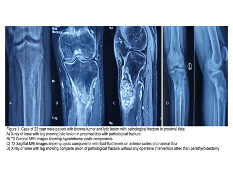
Five out of our six patients were females. Also 5 out of our 6 patients were under the age of 25. 2 patients presented to us with multiple lytic lesions and other 4 patients had solitary lesions. 1 patient out of 6 presented to us with pathological fracture. Only 2 out of the 6 patients had a positive sestamibi scan but Ultrasonography (USG) of the neck and MRI of the neck showed the presence of parathyroid adenoma in all the cases. Preoperative average of VAS score in this patients was 67 which was decreased to as low as 24. We performed paired t-tests on the blood investigation values and Visual Analog Scale (VAS) scores for all patients. The results were statistically significant, with values returning to normal three months post-surgery.
Conclusion
When radiographic evidence of a lytic lesion and hypercalcemia are present, Brown tumor should always be considered in the differential diagnosis. Brown tumor has a distinctive imaging appearance, with solid components displaying intermediate to low intensity on T1- and T2-weighted images, while the cystic components appear hyperintense on T2-weighted images and exhibit fluid-fluid levels. MRI of the neck corroborates the ultrasound findings and detects parathyroid adenomas as hyperintense nodes on T2-weighted images. Parathyroidectomy yields excellent results, enabling conservative management of lytic lesions.
References
Chami, B., et al., Brown tumor of the palate as first manifestation of primary hyperparathyroidism: a case report, Med. Buccale Chir. Buccale 17 (2011) 287–291. https://doi.org/10.1051/mbcb/2011136
Silverberg SJ, Bilezikian JP. "Asymptomatic primary hyperparathyroidism: a medical perspective." In: Bilezikian JP, Marcus R, Levine MA, editors. The Parathyroids: Basic and Clinical Concepts. 3rd edition. Amsterdam: Elsevier; 2015. p. 315-330. https://doi.org/10.1016/j.suc.2004.03.002
Townsend CM Jr, Beauchamp RD, Evers BM, et al. Sabiston textbook of surgery.19th ed. Philadel-phia: Elsevier, 2012, p.933. https://books.google.ch/books/about/Sabiston_Textbook_of_Surgery_E_Book.html?id=nvz_mItQyM4C&redir_esc=y
Costanzo L S. BRS physiology. 4th ed.Philadelphia: Lippincott Williams Wilkins,2011, pp.247–248. https://pnashr.pub/wp-content/uploads/2020/07/BRS-Physiology.pdf
Samarendra MS, Kriplani D, Rajendran J, Maheshwari M. Brown Tumor of Hyperparathyroidism Mimicking Metastatic Bone Disease: A Diagnostic Dilemma. Indian Journal of Endocrinology and Metabolism. 2013;17 (Suppl 1):S239-S241.
Bilezikian JP, Brandi ML, Eastell R, Silverberg SJ, Udelsman R, Marcocci C, Potts JT Jr. Guidelines for the management of asymptomatic primary hyperparathyroidism: summary statement from the Fourth International Workshop. The Journal of Clinical Endocrinology & Metabolism. 2014;99(10):3561-3569. https://doi.org/10.1210/jc.2014-1413
Zanocco KA, Yeh MW. Primary Hyperparathyroidism: Effects on Bone Health. Endocrinol Metab Clin North Am. 2017 Mar;46(1):87–104. https://doi.org/10.1016/j.ecl.2016.09.012
Younes NA, Shafagoj Y, Khatib F, Ababneh M. Laboratory screening for hyperparathyroidism. Clin Chim Acta. 2005 Mar;353(1–2):1–12. https://doi.org/10.1016/j.cccn.2004.10.003
Fraser WD. Hyperparathyroidism. Lancet. 2009;374 (9684):145–58. https://doi.org/10.1016/S0140-6736(09)60507-9
Choi JH, Kim KJ, Lee YJ, Kim SH, Kim SG, Jung KY, Choi DS, Kim NH. Primary Hyperparathyroidism with Extensive Brown Tumors and Multiple Fractures in a 20-Year-Old Woman. Endocrinol Metab (Seoul). 2015 Dec;30(4):614–9. https://doi.org/10.3803/EnM.2015.30.4.614
Pappu R, Jabbour SA, Reginato AM, Reginato AJ. Musculoskeletal manifestations of primary hyperparathyroidism. Clin Rheumatol. 2016 Dec;35(12):3081–7. https://doi.org/10.1007/s10067-016-3450-3
Keyser JS, Postma GN. Brown tumor of the mandible. Am J Otolaryngol.1996 Nov-Dec;17(6):407–10. https://doi.org/10.1016/S0196-0709(96)90075-7
J. Liu, N.E. Cusano, B.C. Silva, L. Zhao, X. He, B. Tao, L. Sun, H. Zhao, W. Fan, M.E. Romano, G. Ning, J.P. Bilezikian, Primary hyperparathyroidism: a tale of two cities revisited. Bone Res. 2, 162–169 (2013). https://doi.org/10.4248/BR201302005
Psaila A, Conti L, Azzopardi AP and Coppini DV: A brown tumor secondary to hyperparathyroidism in the maxilla, skull, scapula, and femora. Proc (Bayl Univ Med Cent) 34(1): 163-165, 2020. https://doi.org/10.1080/08998280.2020.1826260
Ngo QX, Ngo DQ, Tran TD, Le DT, Hoang GN and Le QV: Multiple brown tumors with primary hyperparathyroidism mimicking bone metastases. Int J Surg Case Rep 79: 375-378, 2021. https://doi.org/10.1016/j.ijscr.2021.01.002
Dhaniwala NS and Dhaniwala MN: Multiple Brown tumors in a case of primary hyperparathyroidism with pathological fracture in femur. J Orthop Case Rep 10(6): 49-53, 2020. https://www.ncbi.nlm.nih.gov/pmc/articles/PMC7815671/
Lenherr-Taube N, Lam CK, Vali R, Shammas A, Campisi P,Zawawi F, Somers GR, Stimec J, Mete O, Wong AK and Sochett E: Severe primary hyperparathyroidism caused by parathyroid carcinoma in a 13-year-old child; novel findings from HRpQCT. JBMR Plus 4(3): e10324, 2020. https://doi.org/10.1002/jbm4.10324
Hu J, He S, Yang J, Ye C, Yang X and Xiao J: Management of brown tumor of spine with primary hyperparathyroidism: A case report and literature review. Medicine (Baltimore) 98(14): e15007, 2019. https://doi.org/10.1097/MD.0000000000015007
Aslan S, Bilgici MC, Bernay RF, Aydin HM and Selcuk MB: Parathyroid adenoma presenting with multiple Brown tumors in an adolescent patient. North Clin Istanb 5(4): 361-364, 2018. https://doi.org/10.14744/nci.2018.35693.
Goyal A, Boro H and Khadgawat R: Brown tumor as an index presentation of severe vitamin D deficiency in a teenage girl. Cureus 10(5): e2722, 2018. https://doi.org/10.7759/cureus.2722.
Panagopoulos A, Tatani I, Kourea HP, Kokkalis ZT, Panagopoulos K and Megas P: Osteolytic lesions (brown tumors) of primary hyperparathyroidism misdiagnosed as multifocal giant cell tumor of the distal ulna and radius: a case report. J Med Case Rep 12(1):176, 2018. https://doi.org/10.1186/s13256-018-1723-y
Garla VV, Akhtar I, Salim S and Subauste A: Osteitis fibrosa cystica masquerading as bone neo-plasm. BMJ Case Rep 2018. https://doi.org/10.1136/bcr-2018-224546
Baig MN, Mac Dhaibheid C and Shannon FJ: Hip fracture in a patient with primary hyperparathyroidism: medical and surgical lessons. Cureus 10(1): e2104, 2018. https://doi.org/10.7759/cureus.2104
Misiorowski W, Czajka-Oraniec I, Kochman M, Zgliczyński W and Bilezikian JP: Osteitis fibrosa cystica - a forgotten radiological feature of primary hyperparathyroidism. Endocrine 58(2): 380-385, 2017. https://doi.org/10.1007/s12020-017-1414-2
Shekhawat VS and Bhansali A: Vanishing metatarsal: a rare manifestation of primary hyperparathyroidism. BMJ Case Rep 2017: bcr2017 220676, 2017. https://doi.org/10.1136/bcr-2017-220676
Vaishya R, Agarwal AK, Vijay V and Vaish A: A brown tumor of tibial diaphysis masquerading as malignancy. Cureus 9(6): e1319, 2017. https://doi.org/10.7759/cureus.1319
Jervis L, James M, Howe W and Richards S: Osteolytic lesions: osteitis fibrosa cystica in the setting of severe primary hyperparathyroidism. BMJ Case Rep 2017. https://doi.org/10.1136/bcr-2017-220603
Schnyder MA, Stolzmann P, Huber GF and Schmid C: A patient with a history of breast cancer and multiple bone lesions: a case report. J Med Case Rep 11(1): 127, 2017. https://doi.org/10.1186/s13256-017-1296-1
Park SH, Kong GM, Kwon YU and Park JH: Pathologic fracture of the femur in brown tumor induced in parathyroid carcinoma: a case report. Hip Pelvis 28(3): 173-177, 2016. https://doi.org/10.5371/hp.2016.28.3.173
Riestra Fernández M, Suárez Gutiérrez L, Martínez M and Diéguez Felechosa M: Primary hyperparathyroidism: An unusual presentation. Reumatol Clin 13(4): 246-247, 2017. https://doi.org/10.1016/j.reuma.2016.07.004
Hussain M and Hammam M: Management challenges with brown tumor of primary hyperparathyroidism masked by severe vitamin D deficiency: a case report. J Med Case Rep 10: 166, 2016. https://doi.org/10.1186/s13256-016-0933-4
Sathyakumar S, Cherian KE, Shetty S and Paul TV: Impact of curative surgery on bone in a patient with osteitis fibrosa cystica of primary hyperparathyroidism. BMJ Case Rep 2016: https://doi.org/10.1136/bcr-2016-214970
Downloads
Published
Issue
Section
License
Copyright (c) 2024 ABHIJEET ASHOK SALUNKE

This work is licensed under a Creative Commons Attribution 4.0 International License.
This license requires that reusers give credit to the creator. It allows reusers to distribute, remix, adapt, and build upon the material in any medium or format, even for commercial purposes.


