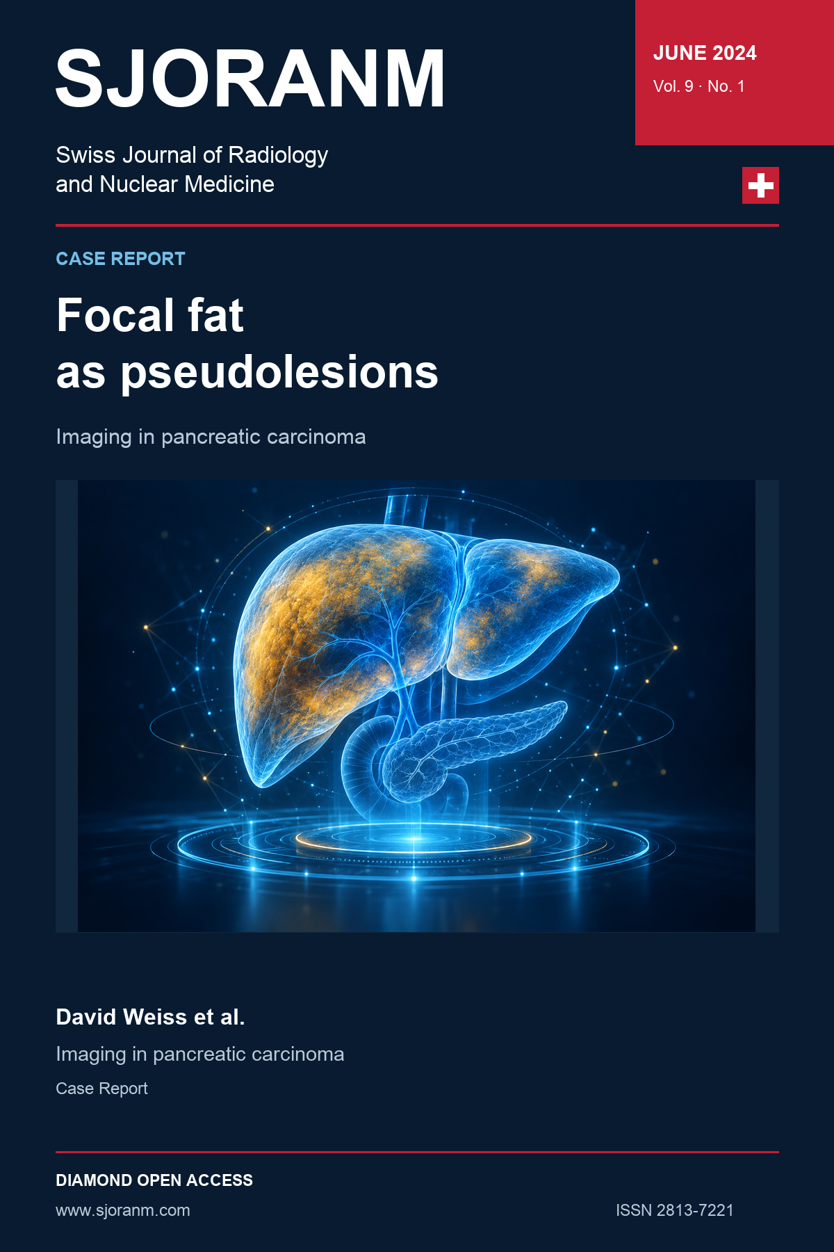
Inhomogeneous Focal Fat Distribution as Pseudolesions in a Patient with Pancreatic Carcinoma
DOI:
https://doi.org/10.59667/sjoranm.v9i1.16Keywords:
hepatic pseudolesion, focal steatosis, focal fat sparing, pancreatic adenocarcinoma, diffusion-weighted imaging, hepatobiliary-phase MRI, contrast-enhanced ultrasonographyAbstract
A 66-year-old patient underwent liver magnetic resonance imaging during initial staging of pancreatic adenocarcinoma. Several focal hepatic abnormalities raised concern for metastases. Their signal characteristics on T1-weighted imaging, absence of corresponding diffusion restriction, preserved hepatobiliary-phase enhancement, and benign appearance on contrast-enhanced ultrasonography supported interpretation as pseudolesions related to heterogeneous hepatic fat distribution rather than metastatic disease. The patient subsequently underwent Whipple surgery. Follow-up magnetic resonance imaging two years later demonstrated locoregional recurrence without hepatic lesions, further supporting the original benign interpretation. This case illustrates how multiparametric magnetic resonance imaging, complementary ultrasonography, and longitudinal follow-up can increase diagnostic confidence when focal fat alteration mimics liver metastases.
References
1. Ramanathan S, Raghu V, Virmani V, Sheikh A, Al Heidous M, Tirumani S. Unveiling the unreal: Comprehensive imaging review of hepatic pseudolesions. Clin Imaging. 2021;80:439-453. https://doi.org/10.1016/j.clinimag.2021.09.008 DOI: https://doi.org/10.1016/j.clinimag.2021.09.008
2. Metin NO, Karaosmanoğlu AD, Metin Y, Karçaaltıncaba M. Focal hypersteatosis: a pseudolesion in patients with liver steatosis. Diagn Interv Radiol. 2019;25(1):14-20. https://doi.org/10.5152/dir.2018.17519 DOI: https://doi.org/10.5152/dir.2018.17519
3. Elsayes KM, Menias CO, Morshid AI, Shaaban AM, Fowler KJ, Tang A, Chernyak V, Szklaruk J, Bashir MR. Spectrum of pitfalls, pseudolesions, and misdiagnoses in noncirrhotic liver. AJR Am J Roentgenol. 2018;211(1):97-108. https://doi.org/10.2214/AJR.18.19820 DOI: https://doi.org/10.2214/AJR.18.19820
4. Fruehwald-Pallamar J, Bastati-Huber N, Fakhrai N, Jantsch M, Puchner S, Herneth AM, Ba-Ssalamah A. Confident non-invasive diagnosis of pseudolesions of the liver using diffusion-weighted imaging at 3T MRI. Eur J Radiol. 2012;81(6):1353-1359. https://doi.org/10.1016/j.ejrad.2011.03.072 DOI: https://doi.org/10.1016/j.ejrad.2011.03.072
5. Donati OF, Fischer MA, Chuck N, Hunziker R, Weishaupt D, Reiner CS. Accuracy and confidence of Gd-EOB-DTPA enhanced MRI and diffusion-weighted imaging alone and in combination for the diagnosis of liver metastases. Eur J Radiol. 2013;82(5):822-828. https://doi.org/10.1016/j.ejrad.2012.12.005 DOI: https://doi.org/10.1016/j.ejrad.2012.12.005
Downloads
Published
Data Availability Statement
The clinical and imaging information supporting this case is contained within the article. Further information may be requested from the corresponding author, subject to patient privacy and applicable institutional requirements.
Issue
Section
License
Copyright (c) 2024 David Weiss, Martin Maurer

This work is licensed under a Creative Commons Attribution 4.0 International License.
This license requires that reusers give credit to the creator. It allows reusers to distribute, remix, adapt, and build upon the material in any medium or format, even for commercial purposes.
 LinkedIn ↗
 X ↗
 VK ↗
 Truth Social ↗
 Bluesky ↗
 YouTube ↗