
Typical and atypical appearance of microwave ablation zones in the liver
DOI:
https://doi.org/10.59667/sjoranm.v2i1.12Keywords:
microwave ablation, post-interventional imaging, HCC, oligo-metastasisAbstract
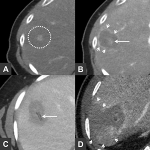
Local minimally invasive interventional procedures such as radiofrequency ablation (RFA) and microwave ablation (MWA) for the treatment of both primary hepatic malignancy and liver metastases of various origins have become increasingly popular in recent years. After treatment, is then the task of follow-up imaging with computed tomography (CT) and/or magnetic resonance imaging (MRI) to evaluate treatment success and to be aware of possible pitfalls that might give rise to misinterpretation.
References
(1) Gao J, Wang SH, Ding XM et al. Radiofrequency ablation for single hepatocellular carcinoma 3 cm or less as first-line treatment. World journal of gastroenterology 2015; 21: 5287-5294
(2) Vogl TJ, Nour-Eldin NA, Hammerstingl RM et al. Microwave Ablation (MWA): Basics, Technique and Results in Primary and Metastatic Liver Neoplasms - Review Article. RoFo : Fortschritte auf dem Gebiete der Rontgenstrahlen und der Nuklearmedizin 2017; 189: 1055-1066
(3) Vogl TJ, Muller PK, Hammerstingl R et al. Malignant liver tumors treated with MR imaging-guided laser-induced thermotherapy: technique and prospective results. Radiology 1995; 196: 257-265
(4) Brace CL. Microwave ablation technology: what every user should know. Current problems in diagnostic radiology 2009; 38: 61-67
(5) Simon CJ, Dupuy DE, Mayo-Smith WW. Microwave ablation: principles and applications. Radiographics : a review publication of the Radiological Society of North America, Inc 2005; 25 Suppl 1: S69-83
(6) Wu H, Exner AA, Krupka TM et al. Radiofrequency ablation: post-ablation assessment using CT perfusion with pharmacological modulation in a rat subcutaneous tumor model. Academic radiology 2009; 16: 321-331
(7) Patel N, King AJ, Breen DJ. Imaging appearances at follow-up after image-guided solid-organ abdominal tumour ablation. Clinical radiology 2017; 72: 680-690
(8) Lencioni R, Cioni D, Bartolozzi C. Percutaneous radiofrequency thermal ablation of liver malignancies: techniques, indications, imaging findings, and clinical results. Abdominal imaging 2001; 26: 345-360
Downloads
Published
Issue
Section
License
Copyright (c) 2023 Martin Maurer

This work is licensed under a Creative Commons Attribution 4.0 International License.
This license requires that reusers give credit to the creator. It allows reusers to distribute, remix, adapt, and build upon the material in any medium or format, even for commercial purposes.


