
The Ultrasound Analysis of the Most Important Musculoskeletal Issues
Visual presentation of common standard ultrasound imaging techniques in the Musculoskeletal area
DOI:
https://doi.org/10.59667/sjoranm.v9i2.12Keywords:
Musculoskeletal ultrasound, MSK sonography, descriptions of the sonographic sections and anatomical structures, definition of standard sonographic section levels, probe positioningAbstract
This work shows the most important standard sections of relevant questions in musculoskeletal (MSK) ultrasound of the wrist, elbow, shoulder, hip, knee and ankle. Exemplary images with descrip-tions of the sections and anatomical structures were created and the positioning of the patient explained. Several patient cases per region were also presented and explained in order to place the described procedures in a clinical context. The advantages and disadvantages of the imaging procedures were explained and compared. MSK ultrasound is particularly strong in the visualisation of superficial structures, whereby this form of imaging is superior to other imaging procedures in some cases. In addition, the detection of inflammation, for example, is not only possible via indirect signs (e.g. free fluid or abscess) compared to computer tomography. In addition, the hyperperfusion caused by inflammation can be visualised directly at the affected site using Doppler examination. Even in the case of functional restrictions such as shoulder impingement or tendon instability (see section 4.6.11: Patient case 19), which are difficult to visualise using other imaging techniques, the tissue can be viewed and moved in vivo using a dynamic examination. This can lead to a better understanding of the pathology and a more precise diagnosis. MSK sonography therefore proves to be a valuable addition to everyday clinical practice.
The focus of our work was the presentation of common standard ultrasound incisions in the MSK area. The intention to keep this guide clear and to cover the common pathologies is limited by the fact that not all sections could be covered. In a future work, further, less frequently used incisions could be shown in order to cover a broader spectrum of possible pathologies. For example, sections of the thorax (such as the thoracolumbar fascia or areas of the spine and lumbar region) could also be shown. A further question is whether MSK ultrasound can save or replace an MRI or CT examination in certain cases. As discussed in the introduction, this would be an advantage from the patient's point of view and in economic terms. Further studies investigating this aspect would be valuable.
References
Sharpe RE, Nazarian LN, Parker L, Rao VM, Levin DC. Dramatically increased musculoskeletal ultrasound utilization from 2000 to 2009, especially by podiatrists in private offices. Journal of the American College of Radiology 2012;9(2):141–6.
Middleton WD, Payne WT, Teefey SA, Hildebolt CF, Rubin DA, Yamaguchi K. Sonography and MRI of the Shoulder: Comparison of Patient Satisfaction. American Journal of Roentgenology [Internet] 2004 [cited 2023 Oct 27];183(5):1449–52. Available from: https://pubmed.ncbi.nlm.nih.gov/15505319/
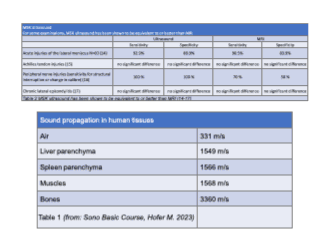
Hofer Matthias. Sono Grundkurs. 11th ed. Stuttgart: Georg Thieme Verlag; 2023.
Narouze Samer N. Atlas of Ultrasound-Guided Procedures in Interventional Pain Management [Internet]. New York, NY: Springer New York; 2011. Available from: http://link.springer.com/10.1007/978-1-4419-1681-5
Nazarian LN. The Top 10 Reasons Musculoskeletal Sonography Is an Important Complementary or Alternative Technique to MRI. American Journal of Roentgenology [Internet] 2008 [cited 2023 Oct 27];190(6):1621–6. Available from: https://www.ajronline.org/doi/10.2214/AJR.07.3385#sec-3
Bangard C, Paszek J, Berg F, et al. MR imaging of claustrophobic patients in an open 1.0T scanner: Motion artifacts and patient acceptability compared with closed bore magnets. Eur J Radiol [Internet] 2007 [cited 2023 Oct 27];64(1):152–7. Available from: https://pubmed.ncbi.nlm.nih.gov/17374468/
Neumann T, Ermert H. Schlieren visualization of ultrasonic wave fields with high spatial resolution. Ultrasonics 2006;44(SUPPL.).
Link TM, Majumdar S, Peterfy C, et al. HIGH RESOLUTION MRI OF SMALL JOINTS: IMPACT OF SPATIAL RESOLUTION ON DIAGNOSTIC PERFORMANCE AND SNR. 1998.
Beggs I, Stefano Bianchi U, Angel Bueno S, et al. European Society of MusculoSkeletal Radiology Musculoskeletal Ultrasound Technical Guidelines I. Shoulder. 2010.
Khoury V, Cardinal É, Bureau NJ. Musculoskeletal Sonography: A Dynamic Tool for Usual and Unusual Disorders. American Journal of Roentgenology [Internet] 2007 [cited 2023 Oct 27];188(1):W63–73. Available from: https://www.ajronline.org/doi/10.2214/AJR.06.0579
Beggs I, Stefano Bianchi U, Angel Bueno S, et al. European Society of MusculoSkeletal Radiology Musculoskeletal Ultrasound Technical Guidelines II. Elbow. 2010.
Neustadter J, Raikin SM, Nazarian LN. Dynamic Sonographic Evaluation of Peroneal Tendon Subluxation. American Journal of Roentgenology [Internet] 2004 [cited 2023 Oct 27];183(4):985–8. Available from: https://pubmed.ncbi.nlm.nih.gov/15385290/
O’Neill J. Musculoskeletal ultrasound: Anatomy and technique. Springer New York; 2008.
Elshimy A, Osman AM, Awad MES, Abdel Aziz MM. Diagnostic accuracy of point-of-care knee ultrasound for evaluation of meniscus and collateral ligaments pathology in comparison with MRI. Acta radiol 2023;64(7):2283–92.
Gatz M, Bode D, Betsch M, et al. Multimodal Ultrasound Versus MRI for the Diagnosis and Monitoring of Achilles Tendinopathy: A Prospective Longitudinal Study. Orthop J Sports Med 2021;9(4):232596712110068.
Nischal N, Gupta S, Lal K, Singh JP. Performance Evaluation of High-Resolution Ultrasound versus Magnetic Resonance Imaging in Diagnosing Peripheral Nerve Pathologies. Indian Journal of Radiology and Imaging 2021;31(01):043–8.
Bachta A, Rowicki K, Kisiel B, et al. Ultrasonography versus magnetic resonance imaging in detecting and grading common extensor tendon tear in chronic lateral epicondylitis. PLoS One 2017;12(7):e0181828.
Murphy A, Morgan M. Musculoskeletal ultrasound [Internet]. Radiopaedia.org. 2014 [cited 2023 Oct 27];Available from: https://radiopaedia.org/articles/31803
Elliot Duff et al. Medical Ultrasound [Internet]. Wikipedia.org. 2002 [cited 2023 Dec 8];Available from: https://en.wikipedia.org/w/index.php?title=Medical_ultrasound&oldid=1182553254
Page P, Manske RC, Voight M, Wolfe C. MSK Ultrasound - An IJSPT Perspective. Int J Sports Phys Ther 2023;18(1).
Stanislavsky A, Patel M. Biceps long head tendon tear [Internet]. Radiopaedia.org. 2019 [cited 2023 Nov 8];Available from: https://radiopaedia.org/cases/65346
Patel M. Lipoma - subdeltoid [Internet]. Radiopaedia.org. 2023 [cited 2023 Nov 8];Available from: https://radiopaedia.org/cases/163404
Jin T, Patel M. Supraspinatous tendon tear [Internet]. Radiopaedia.org. 2010 [cited 2023 Nov 8];Available from: https://radiopaedia.org/cases/12576
Botz B. High grade rupture of the distal triceps tendon [Internet]. Radiopaedia.org. 2022 [cited 2023 Nov 6];Available from: https://radiopaedia.org/cases/98612
Patel M. Anconeus epitrochlearis [Internet]. Radiopaedia.org. 2020 [cited 2023 Nov 6];Available from: https://radiopaedia.org/cases/84506
Patel M. Medial elbow snapping [Internet]. Radiopaedia.org. 2023 [cited 2023 Nov 6];Available from: https://radiopaedia.org/cases/166258
Beggs I, Stefano Bianchi U, Angel Bueno S, et al. European Society of MusculoSkeletal Radiology Musculoskeletal Ultrasound Technical Guidelines III. Wrist. 2010.
Patel M. Wrist laceration (ultrasound) [Internet]. Radiopaedia.org. 2021 [cited 2023 Nov 6];Available from: https://radiopaedia.org/cases/86723
Patel M. Carpal tunnel syndrome [Internet]. Radiopaedia.org. 2018 [cited 2023 Nov 8];Available from: https://radiopaedia.org/cases/63771
Patel M. Scaphotrapezial joint ganglion cyst [Internet]. Radiopaedia.org. 2021 [cited 2023 Nov 9];Available from: https://radiopaedia.org/cases/95326
Beggs I, Stefano Bianchi U, Angel Bueno S, et al. European Society of MusculoSkeletal Radiology Musculoskeletal Ultrasound Technical Guidelines IV. Hip. 2010.
Normal Ultrasound Anatomy of the Musculoskeletal System. Springer Milan; 2012.
Wu WT, Chang KV, Lin CP, Yeh CC, Özçakar L. Ultrasound imaging for inguinal hernia: a pictorial review. Ultrasonography 2022;41(3):610–23.
Patel M. Adductor longus tear [Internet]. Radiopaedia.org. 2021 [cited 2023 Nov 9];Available from: https://radiopaedia.org/cases/90111
Jones J, Cid Barría L. Hip joint septic arthritis (ultrasound) [Internet]. Radiopaedia.org. 2021 [cited 2023 Nov 9];Available from: https://radiopaedia.org/cases/86091
Alkhateeb K. Acetabular labral tear hip [Internet]. Radiopaedia.org. 2019 [cited 2023 Nov 16];Available from: https://radiopaedia.org/cases/72216
Petersilge CA. Chronic Adult Hip Pain: MR Arthrography of the Hip. RadioGraphics 2000;20(suppl_1):S43–52.
Chmiel-Nowak M, Schubert R. Acetabular labral tear [Internet]. Radiopaedia.org. 2011 [cited 2023 Nov 16];Available from: https://radiopaedia.org/articles/14047
Beggs I, Stefano Bianchi U, Angel Bueno S, et al. European Society of MusculoSkeletal Radiology Musculoskeletal Ultrasound Technical Guidelines V. Knee. 2010.
Patel M. Quadriceps tendon tear [Internet]. Radiopaedia.org. 2011 [cited 2023 Nov 9];Available from: https://radiopaedia.org/cases/12805
Bickle I, Patel M. Tenosynovial giant cell tumour (knee) [Internet]. Radiopaedia.org. 2019 [cited 2023 Nov 9];Available from: https://radiopaedia.org/cases/71989
Patel M. Deep infrapatellar bursitis [Internet]. Radiopaedia.org. 2021 [cited 2023 Nov 9];Available from: https://radiopaedia.org/cases/86278
Beggs I, Stefano Bianchi U, Angel Bueno S, et al. European Society of MusculoSkeletal Radiology Musculoskeletal Ultrasound Technical Guidelines VI. Ankle. 2010.
Patel M. Anterior talofibular ligament injury [Internet]. Radiopaedia.org. 2019 [cited 2023 Nov 9];Available from: https://radiopaedia.org/cases/66965
Patel M. Achilles tendon tear [Internet]. Radiopaedia.org. 2020 [cited 2023 Nov 9];Available from: https://radiopaedia.org/cases/76966
Patel M. Peroneus brevis tear [Internet]. Radiopaedia.org. 2023 [cited 2023 Nov 9];Available from: https://radiopaedia.org/cases/168567
Bureau NJ, Ziegler D. Economics of Musculoskeletal Ultrasound. Curr Radiol Rep. 2016;4(8).
Downloads
Published
Issue
Section
License
Copyright (c) 2024 Dan Tschanz, Paolo Lombardo

This work is licensed under a Creative Commons Attribution 4.0 International License.
This license requires that reusers give credit to the creator. It allows reusers to distribute, remix, adapt, and build upon the material in any medium or format, even for commercial purposes.


