The Magnetic Resonance Imaging of a Rare Case of Primary Choriocarcinoma of the Vulva
DOI:
https://doi.org/10.59667/sjoranm.v9i1.12Keywords:
Gestational Choriocarcinoma, Vulva, Gestational Trophoblastic Neoplasia (GTN), Magnetic Resonance Imaging (MRI)Abstract
Primary extrauterine choriocarcinoma is a rare condition, commonly reported in the cervix. Primary vulvovaginal choriocarcinoma has rarely been reported and review of literature reveals no case with MRI features for choriocarcinoma involving only the vulva. We report a rare case of primary choriocarcinoma of the vulva in a young woman, highlighting its Magnetic Resonance Imaging (MRI) features.
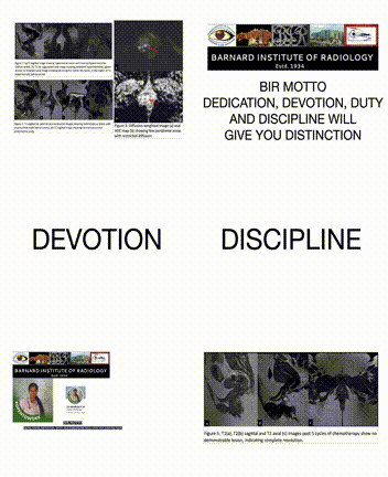
A 26-year-old female patient presented with complaints of menorrhagia, elevated serum Beta human chorionic gonadotropin (β-hCG) levels and a history of suction evacuation for partial molar pregnancy 4 months ago. Magnetic Resonance Imaging (MRI) of the pelvis revealed a normal uterus and adnexa. However, there was a well-defined T1 hypointense, T2 heterogenously hyperintense sublabial mass lesion with internal flow voids, few peripheral areas of restricted diffusion and few internal areas of T1 hyperintensity that bloomed on gradient images. On contrast administration, there was intense peripheral enhancement of the lesion, especially in the regions of restricted diffusion and also in the center of the lesion, where flow voids were observed. Few confluent non enhancing areas of necrosis were also present. In view of the clinical presentation, elevated β-hCG and imaging findings, a diagnosis of choriocarcinoma of the vulva was made and chemotherapy (EMA-CO regimen) was initiated, following which β-hCG decreased progressively and the patient is currently asymptomatic and follow up MRI revealed no abnormal significant residual lesion.
Awareness of a labial lesion being due to extrauterine choriocarcinoma in patients with elevated β-hCG is crucial for effective management and recovery. Our case uniquely emphasizes pretreatment MRI features as well as post-treatment imaging follow-up
References
KAIRI-VASSILATOU E, PAPAKONSTANTINOU K, GRAPSA D, KONDI-PAPHITI A, HASIAKOS D. Primary gestational choriocarcinoma of the uterine cervix. report of a case and review of the literature. International Journal of Gynecological Cancer. 2007 Jul;17(4):921–5. doi:10.1111/j.1525-1438.2006.00852.x
Yahata T, Kodama S, Kase H, Sekizuka N, Kurabayashi T, Aoki Y, et al. Primary choriocarcinoma of the uterine cervix: Clinical, MRI, and color Doppler ultrasonographic study. Gynecologic Oncology. 1997 Feb;64(2):274–8. doi:10.1006/gyno.1996.4541
Weiss S, Amit A, Schwartz MR, Kaplan AL. Primary choriocarcinoma of the Vulva. International Journal of Gynecological Cancer. 2001 May 8;11(3):251–4. doi:10.1046/j.1525-1438.2001.01005.x
Boufettal H. Vulva choriocarcinoma. Pan African Medical Journal. 2016;24. doi:10.11604/pamj.2016.24.328.10482
Bhattacharyya SK, Saha SP, Mukherjee G, Samanta J. Metastatic vulvo-vaginal choriocarcinoma mimicking a bartholin cyst and vulvar hematoma-two unusual presentations. Journal of the Turkish German Gynecological Association. 2012 Sept 1;13(3):218–20. doi:10.5152/jtgga.2011.73
Esen R, Mammadzada N, Yildiz B, Karadurmus N. Primary vulvovaginal choriocarcinoma patient who underwent autologous hematopoetic stem cell transplantation: A case report of unusual presentation. Eastern Journal of Medicine. 2018;23(4):347–9. doi:10.5505/ejm.2018.95866
Prakash A, Kumar I, Rajan M, Verma A. MRI of rare vaginal tumors: Report of two cases. Case Reports in Clinical Radiology. 2023 Apr 12;1:80–3. doi:10.25259/crcr_6_2023
Wong T, Fung EP, Yung AW. Primary gestational choriocarcinoma of the vagina: Magnetic Resonance Imaging findings. Hong Kong Medical Journal. 2016 Apr 13;181–3. doi:10.12809/hkmj144362
Allen SD, Lim AK, Seckl MJ, Blunt DM, Mitchell AW. Radiology of gestational trophoblastic Neoplasia. Clinical Radiology. 2006 Apr;61(4):301–13. doi:10.1016/j.crad.2005.12.003
Yingna S, Yang X, Xiuyu Y, Hongzhao S. Clinical characteristics and treatment of gestational trophoblastic tumor with vaginal metastasis. Gynecologic Oncology. 2002 Mar;84(3):416–9. doi:10.1006/gyno.2001.6540

Downloads
Published
Issue
Section
License
Copyright (c) 2024 Prof. Dr. Babu P. Sathyanathan, Venkatakrishnamurali Maheswari Mridhula

This work is licensed under a Creative Commons Attribution 4.0 International License.
This license requires that reusers give credit to the creator. It allows reusers to distribute, remix, adapt, and build upon the material in any medium or format, even for commercial purposes.


