The Utility of multimodality imaging in a curious case of crippled child
Classical type of Trevor's disease
DOI:
https://doi.org/10.59667/sjoranm.v9i1.14Keywords:
Dysplasia epiphysealis hemimelica, Trevor's disease, osteochondromaAbstract
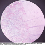
The aim of the article is to establish the importance of devising and adhering to an imaging protocol for musculoskeletal imaging by presenting a case of Dysplasia epiphysealis hemimelica also known as Trevor’s disease in a 13 year old child with atypical presentation which proved to be a diagnostic challenge.
References
Trevor D. Tarso-epiphysial aclasis; a congenital error of epiphysial development. J Bone Joint Surg Br 1950; 32-B:204–213
Fairbank TJ. Dysplasia epiphysialis hemimelica (tarso-ephiphysial aclasis). J Bone Joint Surg Br 1956; 38:237–257
Smith EL, Raney EM, Matzkin EG, Fillman RR, Yandow SM. Trevor's disease: the clinical manifestations and treatment of dysplasia epiphysealis hemimelica. J Pediatr Orthop B 2007; 16:297–302
Arealis G, Nikolaou VS, Lacon A, Ashwood N, Hayward K, Karagkevrekis C. Trevor's disease: a literature review regarding classification, treatment, and prognosis apropos of a case. Case Rep Orthop 2014; 2014:940360
Shinozaki T, Ohfuchi T, Watanabe H, Aoki J, Fukuda T, Takagishi K. Dysplasia epiphysealis hemimelica of the proximal tibia showing epiphy-seal osteochondroma in an adult. Clin Imaging 1999; 23:168–171
Gökkus K, Atmaca H, Sagtas E, Saylik M, Aydin AT. Trevor's disease: up-to-date review of the literature with case series. J Pediatr Orthop B 2017; 26:532–545

Downloads
Published
Issue
Section
License
Copyright (c) 2024 Nandhini Gopi Bagya, Babu Peter Sathyanathan

This work is licensed under a Creative Commons Attribution 4.0 International License.
This license requires that reusers give credit to the creator. It allows reusers to distribute, remix, adapt, and build upon the material in any medium or format, even for commercial purposes.


