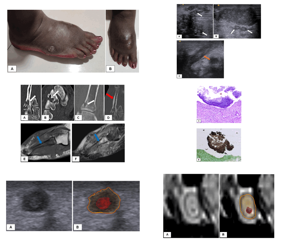
A Radiologist's view on Pedal Actinomycetoma
A Case Report with Comprehensive Review of Literature
DOI:
https://doi.org/10.59667/sjoranm.v21i1.26Keywords:
Pedal mycetoma, Madura foot, Actinomycetoma, Dot-in-circle signAbstract
Background: Tropical diseases comprise of an array of communicable and non-communicable diseases that prevail in the tropical belt. Madura foot, classified as a tropical disease by WHO, is a chronic granulomatous disease that predominantly involves the skin and subcutaneous tissue, commonly affecting the lower limbs. We present a case of actinomycetoma with extensive review of the existing literature, focusing on diagnostic imaging.
Case presentation: A 36-year-old female from eastern India presented with a six-month history of right foot swelling and a discharging wound. She was unsuccessfully treated with multiple courses of antibiotics in local hospitals. Upon referral, radiological investigations were performed for further evaluation. USG showed infiltrative hypoechoic soft tissue with nodular lesions showing targetoid appearance. MRI revealed infiltrative soft tissue with variable sized nodular lesion showing characteristic ‘dot-in-circle' appearance, prompting the diagnosis of pedal mycetoma. Actinomycetoma was confirmed on biopsy.
Conclusions: Pedal mycetoma presents significant diagnostic and therapeutic challenges owing to its insidious progression and delayed diagnosis. Radiological imaging, particularly MRI, plays a pivotal role in diagnosis and staging of the disease, enabling detailed evaluation of soft tissue and bone involvement. The ‘dot-in-circle' sign observed on imaging is pathognomic and aids in accurate diagnosis. Early diagnosis facilitated by diagnostic imaging warrants improved therapeutic outcomes.
References
1. Zumla A, Ustianowski A. Tropical diseases: definition, geographic distribution, transmission, and classification. Infect Dis Clin North Am. 2012; 26: 195-205. https://doi.org/10.1016/j.idc.2012.02.007
2. Cook G.C., Zumla A., editors. Manson's tropical diseases. 22nd edition. Saunders; London: 2009. p. 1830. https://books.google.ch/books?hl=de&lr=&id=CF2INI0O6l0C&oi=fnd&pg=PP1&dq=Cook+G.C.,+Zumla+A.,+editors.+Manson's+tropical+diseases.+22nd+edition.+Saunders;+London:+2009.+p.+1830&ots=N366_HgVBA&sig=8GQfyL71GTuopN5QyM3pRLOkyQ4&redir_esc=y#v=onepage&q&f=false
3. Pierre-Jerome C, Kettner WN. The essentials of Charcot neuropathy. Elsevier; Amsterdam: 2022. P. 223-59. https://books.google.de/books?hl=de&lr=&id=24NTEAAAQBAJ&oi=fnd&pg=PP1&dq=The+essentials+of+Charcot+neuropathy.+Elsevier;+Amsterdam&ots=xS1UCgkonR&sig=RGADYcGUA789TNgNftBDrDt_6lM#v=onepage&q&f=false
4. Karrakchou B, Boubnane I, Senouci K et al. Madurella mycetomatis infection of the foot: a case report of a neglected tropical disease in a non-endemic region. BMC Dermatol. 2020; 20: 1. https://doi.org/10.1186/s12895-019-0097-1
5. Siddig EE, Nyuykonge B, Mhmoud NA, et al. Comparing the performance of the common used eumycetoma diagnostic tests. Mycoses. 2023;66:420-29. https://doi.org/10.1111/myc.13561
6. Carroll DS. Mycetoma pedis. Radiology. 1949; 53: 81–4. https://doi.org/10.1148/53.1.81
7. Cherian RS, Betty M, Manipadam MT, et al. The “dot-in-circle” sign—a characteristic MRI finding in mycetoma foot: a report of three cases. Br J Radiol. 2009; 82: 662–5. https://doi.org/10.1259/bjr/62386689
8. Fahal AH, Shaheen S, Jones DHA. The orthopaedic aspects of mycetoma. Bone Joint J. 2014; 96-B: 420–5. https://doi.org/10.1302/0301-620X.96B3.31421
9. Venkatswami S, Sankarasubramanian A, Subramanyam S. The madura foot: looking deep. Int J Low Extrem Wounds. 2012; 11: 31-42. https://doi.org/10.1177/1534734612438549
10. Czechowski J, Nork M, Haas D et al. MR and other imaging methods in the investigation of mycetomas. Acta Radiologica. 2001; 42: 24-6. https://doi.org/10.1034/j.1600-0455.2001.042001024.x
11. Hailemariam T, Tamiru R, Manyazewal T et al. Madura foot and a continued diagnostic enigma: Dot-in-circle sign on magnetic resonance imaging and ultrasound. IDCases. 2023; 33: e01857. https://doi.org/10.1016/j.idcr.2023.e01857
12. Bentaleb D, Mahdar I, Noureddine L et al. Diagnostic imaging of foot mycetomas: A report on two cases. Radiol Case Rep. 2022; 17: 1817- 23. https://doi.org/10.1016/j.radcr.2022.02.081
13. Cherian RS, Betty M, Manipadam MT et al. The "dot-in-circle" sign -- a characteristic MRI finding in mycetoma foot: a report of three cases. Br J Radiol. 2009; 82: 662-5. https://doi.org/10.1259/bjr/62386689
14. Nabih OO, Bouardi EN, Haloua M et al. Case report: The dot in circle sign: A pathognomonic MRI sign of madura foot. Radiol Case Rep. 2023; 18: 3849- 52. https://doi.org/10.1016/j.radcr.2023.07.052
15. Yadav T, Meena VK, Shaikh M et al. Clinico-radiological-pathological correlation in eumycetoma spectrum: Case series. North Clin Istanb. 2019; 7: 400-6. https://doi.org/10.14744/nci.2019.98215
16. Sarris I, Berendt AR, Athanasous N, et al. MRI of mycetoma of the foot: two cases demonstrating the dot-in-circle sign. Skeletal Radiol. 2003; 32: 179-83. https://doi.org/10.1007/s00256-002-0600-2
17. Fahal AH, Sheik HE, Homeida MM et al. Ultrasonographic imaging of mycetoma. Br J Surg. 1997; 84: 1120-2. https://pubmed.ncbi.nlm.nih.gov/9278658/
18. Ispoglou SS, Zormpala A, Androulaki A et al. Madura foot due to Actinomadura madurae: imaging appearance. J Clin Imaging. 2003; 27: 233-35. https://doi.org/10.1016/S0899-7071(02)00502-8
19. Jimenez AL, Salvo NL. Mycetoma or synovial sarcoma? A case report with review of the literature. J Foot Ankle Surg. 2011; 50: 569-76. https://doi.org/10.1053/j.jfas.2011.04.014
20. Lewall DB, Ofole S, Bendl B. Mycetoma. Skeletal Radiol. 1985;14: 257–62. https://link.springer.com/article/10.1007/BF00352615
21. Jain V, Makwana EG, Bahri N et al. The “Dot in Circle” Sign on MRI in Maduramycosis: A Characteristic Finding. J Clin Imaging Sci. 2012; 2: 66. https://doi.org/10.4103/2156-7514.103056
22. Asly M, Rafaoui A, Bouyermane H, Hakam K, Moustamsik B, Lmidmani F, Rafai M, Largab A, Elfatimi A. Mycetoma [Madura foot]: A case report. Ann Phys Rehabil Med. 2010; 53: 650-4. https://doi.org/10.1016/j.rehab.2010.10.003
23. Serfaty A, Righetti Vieira Ferreira de Araújo A, Severo A et al. Long-term radiographic features of Madura foot. Joint Bone Spine. 2020; 87:167. https://doi.org/10.1016/j.jbspin.2019.09.007
24. Tarafdar S, Kanimozhi P, Sabarish S et al. Magnetic Resonance Imaging in the Diagnosis of Mycetoma with Equivocal Clinical and Laboratory Features. Indian J Dermatol. 2022; 67: 459-463. https://doi.org/10.4103/ijd.ijd_124_21
25. Sidhu R, Patel P, Parikh U et al. Madura foot. Eurorad. 2017; 14374. https://www.researchgate.net/profile/Roopkamal-Sidhu/publication/374367808_Madura_Foot/links/6519c19bb0df2f20a203a57a/Madura-Foot.pdf
26. Elmaataoui A, Elmoustachi A, Aoufi S et al. Eumycetoma due to Madurella mycetomatis from two cases of black grain mycetoma in Morocco. J Mycol Med. 2011; 21: 281- 84. https://doi.org/10.1016/j.mycmed.2011.09.001
27. Brufman T, Ben-Ami R, Mizrahi M et al. Mycetoma of the Foot Caused by Madurella Mycetomatis in Immigrants from Sudan. Isr Med Assoc J. 2015; 17: 418-20. https://europepmc.org/article/med/26357716
28. Martinez EI, Fuentes RC, Sanchez MA et al. Mycetoma foot. Eurorad. 2016; 13444. https://doi.org/10.21203/rs.3.rs-4358839/v1
29. Hoogervorst LA, Op de Coul LS, Ray A, et al. Mycetoma caused by Madurella mycetomatis in immunocompromised patients - a case report and systematic literature review. J Bone Jt Infect. 2022; 7: 241-8. https://doi.org/10.5194/jbji-7-241-2022
30. Gameraddin M, Gareeballah A, Mokhtar S, M Abuzaid M, Alhazmi F, Ali Hamad H. Characterization of Foot Mycetoma Using Sonography and Color Doppler Imaging. Pak J Biol Sci. 2020; 23:968-972. https://doi.org/10.3923/pjbs.2020.968.972
31. Abd El Bagi ME. New radiographic classification of bone involvement in pedal mycetoma. AJR Am J Roentgenol. 2003; 180: 665-8. https://doi.org/10.2214/ajr.180.3.1800665
32. El Shamy ME, Fahal AH, Shakir MY et al. New MRI grading system for the diagnosis and management of mycetoma. Trans R Soc Trop Med Hyg. 2012; 106: 738-42. https://doi.org/10.1016/j.trstmh.2012.08.009
33. Agarwal P, Jagati A, Rathod SP et al. Clinical Features of Mycetoma and the Appropriate Treatment Options. Res Rep Trop Med. 2021; 12: 173-9. https://doi.org/10.2147/RRTM.S282266
34. Grover S, Roy P, Singh G. MADURA FOOT. Med J Armed Forces India. 2001; 57: 163-4. https://doi.org/10.1016/S0377-1237(01)80144-1
35. Relhan V, Mahajan K, Agarwal P et al. Mycetoma: An Update. Indian J Dermatol. 2017; 62: 332-40. https://doi.org/10.4103/ijd.IJD_476_16
36. Salim AO, Mwita CC, Gwer S. Treatment of Madura foot: a systematic review. JBI Database System Rev Implement Rep. 2018; 16: 1519-36. https://doi.org/10.11124/JBISRIR-2017-003433
37. Scolding P, Fahal A, Yotsu RR. Drug therapy for Mycetoma. Cochrane Database Syst Rev. 2018; 2018: CD013082. https://doi.org/10.1002/14651858.CD013082
38. Wadal A, Elhassan TA, Zein HA et al. Predictors of Post-operative Mycetoma Recurrence Using Machine-Learning Algorithms: The Mycetoma Research Center Experience. PLoS Negl Trop Dis. 2016;10: e0005007. https://doi.org/10.1371/journal.pntd.0005007
39. Suleiman SH, Wadaella el S, Fahal AH. The Surgical Treatment of Mycetoma. PLoS Negl Trop Dis. 2016;10: e0004690. https://doi.org/10.1371/journal.pntd.0004690
40. Salamon ML, Lee JH, Pinney SJ. Madura foot [madurella mycetoma] presenting as a plantar fibroma: a case report. Foot Ankle Int. 2006; 27: 212-5. https://doi.org/10.1177/107110070602700311
41. Laohawiriyakamol T, Tanutit P, Kanjanapradit K et al. The "dot-in-circle" sign in musculoskeletal mycetoma on magnetic resonance imaging and ultrasonography. Springerplus. 2014; 3: 671. https://doi.org/10.1186/2193-1801-3-671

Downloads
Published
Data Availability Statement
Data sharing is not applicable to this research article as no new data were created or analysed in this study.
Issue
Section
License
Copyright (c) 2025 Mohan Venugopal, Ebinesh Arulnathan, Bhuvanamha Devi, Poonkodi Manohar, Praislin Gideon

This work is licensed under a Creative Commons Attribution 4.0 International License.
This license requires that reusers give credit to the creator. It allows reusers to distribute, remix, adapt, and build upon the material in any medium or format, even for commercial purposes.


