
A Must-Know Guide to Complex Anal Fistulas: Supralevator, Extrasphincteric, and More
DOI:
https://doi.org/10.59667/sjoranm.v21i1.14Keywords:
EAS: External Anal Sphincter, IAS: Internal Anal Sphincter, VAAFT: video-assisted anal fistula treatment , TROPIS Transanal opening of intersphincteric space , LIFT: ligation of the intersphincteric fistula tract , CLM: Conjoined Longitudinal MuscleAbstract
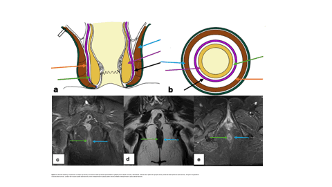
Treatment of perianal fistulae, especially high-grade Grade 5 perianal fistula, is still a difficult procedure in colorectal surgery. Imaging is central to the correct classification, diagnosis, and preoperative evaluation of fistulae. The purpose of this article is to briefly present the five types of perianal fistula with Grade 5 complexity according to the radiological aspects, classification, and therapeutic implications.
References
1) Parks AG, Gordon PH, Hardcastle JD. A classification of fistula-in-ano. Br J Surg. 63 (1):1–12, (1976). https://www.taylorfrancis.com/chapters/edit/10.1201/9780429285714-58/classification-fistula-ano-ana-otero-piñeiro-tracy-hull
2) Halligan, S.J., Magnetic resonance imaging of fistula-in-ano: STIR or SPIR?, British Journal of Radio-logy, Volume 71, Issue 842, 1 February (1998), Pages 141–145. https://doi.org/10.1259/bjr.71.842.9579177
3) Jaime de Miguel Criado, Laura García del Salto, Patricia Fraga Rivas, Luis Felipe Aguilera del Hoyo, Leticia Gutiérrez Velasco, M. Isabel Díez Pérez de las Vacas, Ana G. Marco Sanz, Marcos Manzano Paradela, and Eduardo Fraile Moreno, MR Imaging Evaluation of Perianal Fistulas: Spectrum of Imaging Features, RadioGraphics (2012) 32:1, 175-194. https://doi.org/10.1148/rg.321115040
4) Spencer J A, et. al. Outcome after surgery for perianal fistula: predictive value of MR imaging. American Journal of Roentgenology (1998) 171:2, 403-406. https://ajronline.org/doi/epdf/10.2214/ajr.171.2.9694464
5) Garg P, Yagnik VD, Dawka S, Kaur B, Menon GR. Guidelines to diagnose and treat perilevator high-5 anal fistulas: Supralevator, suprasphincteric, extrasphincteric, high outersphincteric, and high intrarectal fistulas. World J Gastroenterol. (2022) Apr 28;28(16):1608-1624. https://doi.org/10.3748/wjg.v28.i16.1608
6) Bhaya, A. K., & Kumar, N. MRI with MR fistulogram for perianal fistula: a successful combination. Clin Gastrointest Magnetom, (2007) 1(1), 56-59. https://cdn0.scrvt.com/39b415fb07de4d9656c7b516d8e2d907/1800000000012830/b47b587dbaa6/fistulogram_1800000000012830.pdf
7) Steve Halligan and Jaap Stoker Imaging of Fistula in Ano, Radiology (2006) 239:1, 18-33. https://doi.org/10.1148/radiol.2391041043
8) Beckingham, I. J., et. al. Prospective evaluation of dynamic contrast enhanced magnetic resonance imaging in the evaluation of fistula in ano British Journal of Surgery, Volume 83, Issue 10, October (1996), Pages 1396–1398. https://doi.org/10.1002/bjs.1800831022
9) Singh, K., et al., Magnetic Resonance Imaging (MRI) Evaluation of Perianal Fistulae with Surgical Correlation J Clin Diagn Res. (2014) Jun 20;8(6):RC01–RC04 https://doi.org/10.7860/JCDR/2014/7328.4417
10) Chiara Villa, Giovanni Pompili, Giuseppe Franceschelli, Alice Munari, Giovanni Radaelli, Giovanni Maconi, Gian Paolo Cornalba, Role of magnetic resonance imaging in evaluation of the activity of perianal Crohn's disease, European Journal of Radiology, Volume 81, Issue 4, (2012), Pages 616-622, ISSN 0720-048X. https://doi.org/10.1016/j.ejrad.2011.01.046
11) Daabis, N., et al. MRI evaluation of perianal fistulas. The Egyptian Journal of Radiology and Nuclear Medicine, Volume 44, Issue 4, December (2013), Pages 705-711. https://doi.org/10.1016/j.ejrnm.2013.09.003
12) Pomerri, F., et al. Radiologic diagnosis of anal fistulas with radio-opaque markers. La Radiologia Medica, 01 Jun (1988), 75(6):632-637. https://europepmc.org/article/med/3387616
13) Emily J. Bubbers , Kyle G. Cologne, Management of Complex Anal Fistulas. Clin Colon Rectal Surg (2016); 29(01): 043-049. https://doi.org/10.1055/s-0035-1570392
14) Torkzad, M.R., Karlbom, U., MRI for assessment of anal fistula. Insights Imaging 1, 62–71 (2010). https://doi.org/10.1007/s13244-010-0022-y
Downloads
Published
Data Availability Statement
All data generated or analyzed during this study are available from the corresponding author upon reasonable request. The datasets supporting the conclusions of this article have been securely archived and can be accessed for further assessment, subject to ethical and legal considerations. This work has been published in the Swiss Journal of Radiology and Nuclear Medicine.
Issue
Section
License
Copyright (c) 2025 Sushila Muthaiya, Babu Peter Sathyanathan

This work is licensed under a Creative Commons Attribution 4.0 International License.
This license requires that reusers give credit to the creator. It allows reusers to distribute, remix, adapt, and build upon the material in any medium or format, even for commercial purposes.


