Type II Abernethy Malformation with Hepatopulmonary Syndrome: Single-Stage Endovascular Occlusion of an SMV–IVC Communication — A Case Report
DOI:
https://doi.org/10.59667/sjoranm.v36i2.14Keywords:
Abernethy malformation, hepatopulmonary syndrome, plug embolisation, congenital portosystemic shuntAbstract
Background: Congenital extrahepatic portosystemic shunts (Abernethy malformations) can be associated with hyperammonemia and hepatopulmonary syndrome (HPS). Endovascular closure requires careful assessment of shunt anatomy and portal venous drainage.
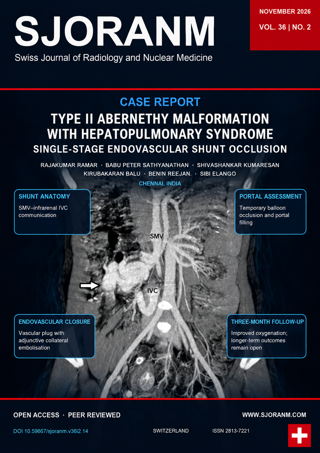
Case presentation: A 12-year-old girl presented with progressive exertional dyspnea, central cyanosis, clubbing, and room-air SpO₂ of 74%. Serum ammonia was 103 µmol/L, while liver biochemistry was within the reference ranges. Echocardiography showed no structural cardiac cause for the cyanosis and agitated saline bubble echocardiography showed delayed appearance (6 cardiac cycles) of bubbles in the left atrium. CT pulmonary angiography demonstrated distal pulmonary vascular dilatation and subpleural telangiectatic vessels, consistent with HPS. The preprocedural ABG on room air showed pH 7.45, PaCO₂ 31 mmHg, PaO₂ 62 mmHg, HCO₃⁻ 22 mEq/L, and A–a oxygen gradient of 49 mmHg; the follow-up ABG normalized. The baseline SpO₂/PaO₂ discrepancy remained unresolved. Abdominal CT angiography showed a side-to-side communication between the superior mesenteric vein (SMV) and infrarenal inferior vena cava (IVC), with tortuous collateral channels and normal, patent intrahepatic portal branches. During temporary balloon occlusion, portal pressure rose from 7 to 17 mmHg, and contrast opacified the intrahepatic portal branches and hepatic parenchyma. The primary communication was occluded with a 12-mm Amplatzer Vascular Plug II, with adjunctive gelfoam embolisation of collateral channels.
Outcome: Doppler ultrasound demonstrated hepatopetal portal flow during early follow-up. At 3 months, room-air SpO₂ was 94%, central cyanosis had resolved, serum ammonia had decreased to 46.8 µmol/L, and the follow-up ABG normalized.
Conclusion: This case highlights the role of cross-sectional vascular mapping and temporary balloon occlusion assessment before single-stage endovascular closure of a congenital portosystemic shunt.
References
1. Ghuman SS, Gupta S, Buxi TB, Rawat KS, Yadav A, Mehta N, et al. The Abernethy malformation—myriad imaging manifestations of a single entity. Indian J Radiol Imaging. 2016;26(3):364–372. https://doi.org/10.4103/0971-3026.190420 DOI: https://doi.org/10.4103/0971-3026.190420
2. McLin VA, Franchi-Abella S, Brütsch T, Bahadori A, Casotti V, de Ville de Goyet J, et al. Expert management of congenital portosystemic shunts and their complications. JHEP Rep. 2024;6(1):100933. https://doi.org/10.1016/j.jhepr.2023.100933 DOI: https://doi.org/10.1016/j.jhepr.2023.100933
3. Kumar P, Bhatia M, Garg A, Jain S, Kumar K. Abernethy malformation: a comprehensive review. Diagn Interv Radiol. 2022;28(1):21–28. https://doi.org/10.5152/dir.2021.20474 DOI: https://doi.org/10.5152/dir.2021.20474
4. Porres-Aguilar M, Altamirano JT, Torre-Delgadillo A, Charlton MR, Duarte-Rojo A. Portopulmonary hypertension and hepatopulmonary syndrome: a clinician-oriented overview. Eur Respir Rev. 2012;21(125):223–233. https://doi.org/10.1183/09059180.00007211 DOI: https://doi.org/10.1183/09059180.00007211
5. Abernethy J. Account of two instances of uncommon formation in the viscera of the human body. Philos Trans R Soc Lond. 1793;83:59–66. https://doi.org/10.1098/rstl.1793.0010 DOI: https://doi.org/10.1098/rstl.1793.0010
6. Morgan G, Superina R. Congenital absence of the portal vein: two cases and a proposed classification system for portasystemic vascular anomalies. J Pediatr Surg. 1994;29(9):1239–1241. https://doi.org/10.1016/0022-3468(94)90812-5 DOI: https://doi.org/10.1016/0022-3468(94)90812-5
7. Kanazawa H, Nosaka S, Miyazaki O, Sakamoto S, Fukuda A, Shigeta T, et al. The classification based on intrahepatic portal system for congenital portosystemic shunts. J Pediatr Surg. 2015;50(4):688–695. https://doi.org/10.1016/j.jpedsurg.2015.01.009 DOI: https://doi.org/10.1016/j.jpedsurg.2015.01.009
8. Baiges A, Turon F, Simón-Talero M, Tasayco S, Bueno J, Zekrini K, et al. Congenital extrahepatic portosystemic shunts (Abernethy malformation): an international observational study. Hepatology. 2020;71(2):658–669. https://doi.org/10.1002/hep.30817 DOI: https://doi.org/10.1002/hep.30817
9. Lautz TB, Tantemsapya N, Rowell E, Superina RA. Management and classification of type II congenital portosystemic shunts. J Pediatr Surg. 2011;46(2):308–314. https://doi.org/10.1016/j.jpedsurg.2010.11.009 DOI: https://doi.org/10.1016/j.jpedsurg.2010.11.009
10. Blanc T, Guerin F, Franchi-Abella S, Jacquemin E, Pariente D, Soubrane O, et al. Congenital portosystemic shunts in children: a new anatomical classification correlated with surgical strategy. Ann Surg. 2014;260(1):188–198. https://doi.org/10.1097/SLA.0000000000000266 DOI: https://doi.org/10.1097/SLA.0000000000000266
11. Agarwal R, Prasad D, Chauhan G, Verma A. Abernethy malformation masquerading as congenital heart disease: a boy with cyanosis, clubbing, and hypoxia. Cureus. 2023;15(1):e33519. https://doi.org/10.7759/cureus.33519 DOI: https://doi.org/10.7759/cureus.33519
12. Bruckheimer E, Dagan T, Atar E, Schwartz M, Kachko L, Superina R, et al. Staged transcatheter treatment of portal hypoplasia and congenital portosystemic shunts in children. Cardiovasc Intervent Radiol. 2013;36(6):1580–1585. https://doi.org/10.1007/s00270-013-0581-7 DOI: https://doi.org/10.1007/s00270-013-0581-7
13. Ponce-Dorrego MD, Hernández-Cabrero T, Garzón-Moll G. Endovascular treatment of congenital portosystemic shunt: a single-center prospective study. Pediatr Gastroenterol Hepatol Nutr. 2022;25(2):147–162. https://doi.org/10.5223/pghn.2022.25.2.147 DOI: https://doi.org/10.5223/pghn.2022.25.2.147
14. Bombardier B, Alli A, Rohr A, Collins Z, Raval K. A case of two shunts in the endovascular treatment of type II Abernethy syndrome. CVIR Endovasc. 2022;5(1):3. https://doi.org/10.1186/s42155-021-00279-7 DOI: https://doi.org/10.1186/s42155-021-00279-7
15. Hatakeyama K, Tozawa T, Noguchi A, Mori N. Long-term follow-up and transcatheter embolisation of extrahepatic congenital portosystemic shunt with shifting hemodynamics. J Clin Imaging Sci. 2025;15:9. https://doi.org/10.25259/JCIS_156_2024 DOI: https://doi.org/10.25259/JCIS_156_2024
Downloads
Published
Data Availability Statement
All data relevant to this case report are contained within the article. No separate research dataset was generated.
Issue
Section
License
Copyright (c) 2026 Rajakumar Ramar, Babu Peter Sathyanathan, Shivashankar Kumaresan, Kirubakaran Balu, Benin Reejan, Sibi Elango

This work is licensed under a Creative Commons Attribution 4.0 International License.
This license requires that reusers give credit to the creator. It allows reusers to distribute, remix, adapt, and build upon the material in any medium or format, even for commercial purposes.
 LinkedIn ↗
 X ↗
 VK ↗
 Truth Social ↗
 Bluesky ↗
 YouTube ↗