Navigating Complexity
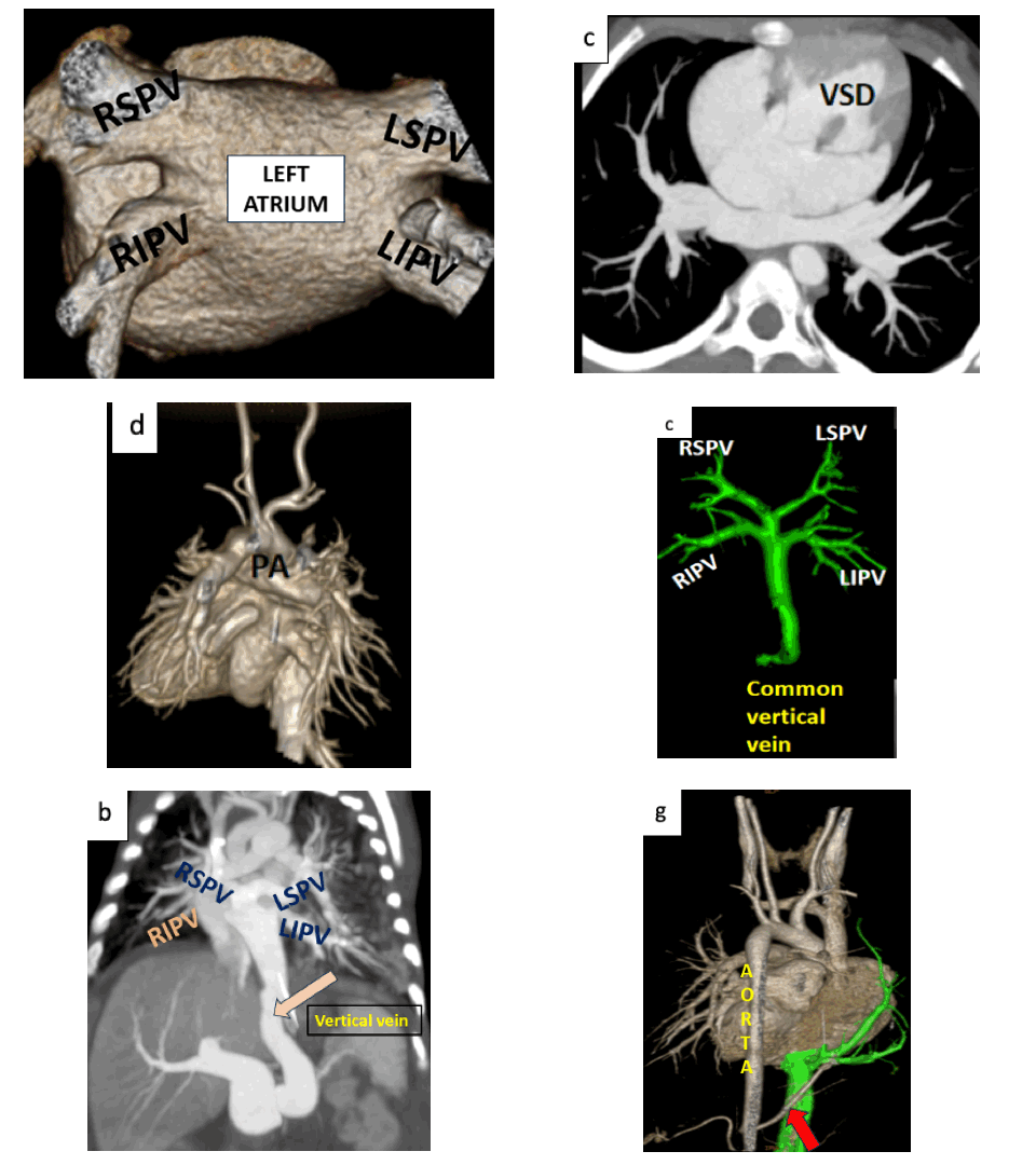
A pictorial representation of anomalous pulmonary venous connection classification in the pediatric population with volume rendering and multiplanar imaging technique
DOI:
https://doi.org/10.59667/sjoranm.v13i1.16Keywords:
TAPVC, volume rendering images, anomalous pulmonary drainage, Scimitar SyndromeAbstract
Anomalous pulmonary venous connections represent a heterogeneous group of congenital heart diseases in which a part or all of the pulmonary veins drains into the right atrium instead of draining into the left atrium. pulmonary veinous anomalies can manifest as partial or total anomalous drainage due to the abnormal embryological development. Multidetector-CT angiography, with its multiplanar reformatting and volume rendering techniques, precisely offers the information about the three-dimensional anatomy and spatial relationships of the cardiovascular structures. Clinical features of anomalous pulmonary venous connections may be silent or have variable features like neonatal cyanosis, volume overload, and pulmonary arterial hypertension due to the left-to-right shunt, which are often associated with other congenital cardiac disease , so that accurate diagnosis is essential for treatment planning
References
1. Lyen, S., Wijesuriya, S., Ngan-Soo, E., et al. Anomalous pulmonary venous drainage: a pictorial essay with a CT focus. J Congenit Heart Dis 1, 7 (2017). https://doi.org/10.1186/s40949-017-0008-4
2. Karamlou T, Gurofsky R, Al Sukhni E, et al. Factors associated with mortality and reoperation in 377 children with total anomalous pulmonary venous connection. Circulation 2007; 115:1591–1598 https://doi.org/10.1161/CIRCULATIONAHA.106.635441
3. White CS, Baffa JM, Haney PJ, Pace ME, and Campbell AB. MR imaging of congenital anomalies of the thoracic veins. RadioGraphics 1997; 17:595–608. https://doi.org/10.1148/radiographics.17.3.9153699
4. CRAIG JM, DARLING RC, ROTHNEY WB. Total pulmonary venous drainage into the right side of the heart; report of 17 autopsied cases not associated with other major cardiovascular anomalies. Lab Invest. 1957 Jan-Feb;6(1):44-64
5. Seale AN, Uemura H, Webber SA, Partridge J, Roughton M, Ho SY, McCarthy KP, Jones S, Shaughnessy L, Sunnegardh J, Hanseus K, Berggren H, Johansson S, Rigby ML, Keeton BR, and Daubeney PE., British Congenital Cardiac Association. Total anomalous pulmonary venous connection: morphology and outcome from an international population-based study. Circulation. 2010 Dec 21;122(25):2718-26. https://doi.org/10.1161/CIRCULATIONAHA.110.940825
6. Berrocal T, Madrid C, Novo S, Gutiérrez J, Arjonilla A, Gómez-Leo N. Congenital anomalies of the tracheobronchial tree, lung, and mediastinum: embryology, radiology, and pathology. RadioGraphics 2004; 24: e17 https://doi.org/10.1148/rg.e17
7. Dillman JR, Yarram SG, and Hernandez RJ. Imaging of pulmonary venous developmental anomalies. Am J Roentgenol. 2009;192(5):1272–85. https://doi.org/10.2214/AJR.08.1526

Downloads
Published
Issue
Section
License
Copyright (c) 2024 Dr. Ramya Selvaraj, Dr. Babu Peter

This work is licensed under a Creative Commons Attribution 4.0 International License.
This license requires that reusers give credit to the creator. It allows reusers to distribute, remix, adapt, and build upon the material in any medium or format, even for commercial purposes.


