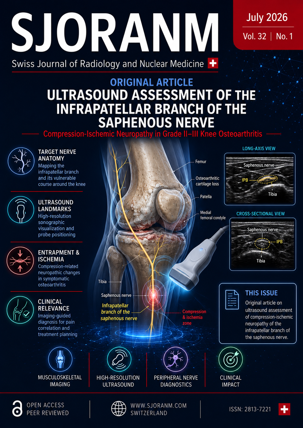
The Ultrasound Verification of Compression Ischemic Neuropathy of the Infrapatellar Nerve in Patients with Grade II–III Knee Osteoarthritis
DOI:
https://doi.org/10.59667/sjoranm.v32i1.14Keywords:
neuropathy, pain syndrome, knee joint, infrapatellar nerve, knee osteoarthritisAbstract
Background / Objective. To perform a detailed ultrasound (US) analysis of pathologically altered periarticular soft tissue structures in the medial compartments of the knee in patients with grade II–III knee osteoarthritis (OA) and to assess the relationship of these changes with anterior medial knee pain.
Materials and Methods. A prospective randomized single‑center study enrolled 82 patients. The main group (n=42) included patients with grade II–III OA, varus/valgus deformity and anterior medial knee pain; the control group (n=40) comprised patients with grade 0–I OA without pain. All participants underwent physical examination (Tinel sign), US assessment of superior femoral ligament (SFL) protrusion and anteroposterior dimension of the interfascial space (APDIFS) in the infrapatellar nerve (IPN) projection, numeric pain rating scale (NRS), and PainDetect questionnaire.
Results. The two groups did not differ significantly in age or sex; women predominated (>98%). SFL protrusion was significantly higher in the main group (7.2–13.8 mm vs. 2.5–6.8 mm, p≤0.05), while APDIFS was significantly lower (0–0.8 mm vs. 1.2–2.1 mm, p≤0.05). Based on SFL protrusion, a practical classification was proposed: type 0 (<7.0 mm) – minimal/no CIN symptoms; type 1 (7.0–10 mm) – episodic symptoms; type 2 (>10 mm) – persistent severe manifestations. Severe CIN was confirmed in 90.4% of type 2 patients (n=38). In 23 type 2 patients not responding to conservative treatment, US‑guided saphenous nerve block and subsequent radiofrequency ablation provided pain regression to 2–3 NRS points in 21 patients; 15 patients underwent total knee arthroplasty with complete pain resolution.
Conclusion. Anterior medial knee pain in OA has a neuropathic origin due to IPN compression caused by SFL protrusion and interfascial space narrowing. US allows reliable verification of these changes, expands clinical interpretation of patient complaints, and helps to choose optimal diagnostic and therapeutic strategies.
References
1. Global Burden of Disease 2021 Osteoarthritis Collaborators. Global, regional, and national burden of osteoarthritis, 1990–2020 and projections to 2050: a systematic analysis for the Global Burden of Disease Study 2021. Lancet Rheumatol. 2023;5(9):e508-e522. DOI: https://doi.org/10.1016/S2665-9913(23)00163-7
2. The Discordance Between Pain and Imaging in Knee Osteoarthritis. J Arthroplasty. 2025;40(8):1992-1999.e1. DOI: https://doi.org/10.1016/j.arth.2025.01.050
3. Ohashi Y, Uchida K, Fukushima K, Inoue G, Takaso M. Mechanisms of Peripheral and Central Sensitization in Osteoarthritis Pain. Cureus. 2023;15(2):e35331. DOI: https://doi.org/10.7759/cureus.35331
4. Perrot S. Pain in osteoarthritis from a symptom to a disease. Best Pract Res Clin Rheumatol. 2023;37(1):101839. DOI: https://doi.org/10.1016/j.berh.2023.101839
5. Miyata T, Kawabata M, Watanabe D, et al. Ultrasound-Guided Manual Therapy for the Infrapatellar Branch of the Saphenous Nerve Entrapment Presenting as Anterior Knee Pain: A Case Report. Cureus. 2025;17(9):e93051. DOI: https://doi.org/10.7759/cureus.93051
6. Drandrov RN, Eldyrev AYu, Nikolaev NS, Kuzmina VA, Andronnikov EA. Method of nerve block with ultrasound navigation for the treatment of pain syndrome in the anterior-medial parts of the knee joint. Russian Federation patent RU 2720157 C1. 2020 Apr 24. (In Russian)
7. Drăghici NC, Bolchis R, Popa LL, Văcăraș V, Iluț S, Bashimov A, Domnița DM, Dragoș HM, Vlad I, Mureșanu DF. Rare entrapment neuropathies of the lower extremity: A narrative review. Medicine (Baltimore). 2024;103(35):e39486. DOI: https://doi.org/10.1097/MD.0000000000039486
8. Zharikova AV, Krivoshey OA, Tsukanova SA. Tunnel neuropathies of the pelvic girdle and lower extremities: practical guide for physicians. Gomel: Republican Scientific and Practical Center for Radiation Medicine and Human Ecology; 2020. (In Russian)
9. Ferre-Martinez A, Miguel-Pérez M, Möller I, Ortiz-Miguel S, Pérez-Bellmunt A, Ruiz N, Sanjuan X, Agullo J, Ortiz-Sagristà J, Martinoli C. Possible Points of Ulnar Nerve Entrapment in the Arm and Forearm: An Ultrasound, Anatomical, and Histological Study. Diagnostics (Basel). 2023;13(7):1332. DOI: https://doi.org/10.3390/diagnostics13071332
10. Kowalska B. Assessment of the utility of ultrasonography with high-frequency transducers in the diagnosis of entrapment neuropathies. J Ultrason. 2014;14(59):371-392. DOI: https://doi.org/10.15557/JoU.2014.0039
11. Mozolevsky YuV, Barinov AN. Complex treatment of tunnel neuropathies of the lower extremities. Neurology, Neuropsychiatry, Psychosomatics. 2013;4:10-21. (In Russian)
12. Drandrov RN, Nikolaev NS, Eldyrev AYu, Andronnikov EA, Kuksova VA. Method for selecting a treatment for compression-ischemic neuropathy of the infrapatellar branch of the saphenous nerve in patients with knee osteoarthritis. Russian Federation patent RU 2799913 C1. 2023 Jul 13. (In Russian)
13. Lyalina VV, Skripnichenko EA, Borisovskaya SV, et al. Neuropathy of the infrapatellar nerve as a cause of chronic pain in the knee joint. Therapy. 2023;9(5(67)):148-155. DOI: https://doi.org/10.18565/therapy.2023.5.148-155 (In Russian)
Downloads
Published
Data Availability Statement
Primary information and the database are provided by the authors upon request
Issue
Section
License
Copyright (c) 2026 MD Evgenii A. Andronnikov, MD Rodion N. Drandrov, MD Alexey Yu. Eldyrev, MD Alexander V. Gogulin

This work is licensed under a Creative Commons Attribution 4.0 International License.
This license requires that reusers give credit to the creator. It allows reusers to distribute, remix, adapt, and build upon the material in any medium or format, even for commercial purposes.