Bilateral precalcaneal congenital fibrolipomatous hamartomas in an eleven-week-old infant: a case report of characteristic ultrasound findings
DOI:
https://doi.org/10.59667/sjoranm.v36i1.14Keywords:
infant, precalcaneal congenital fibrolipomatous hamartoma, plantar nodule, ultrasonography, case reportAbstract
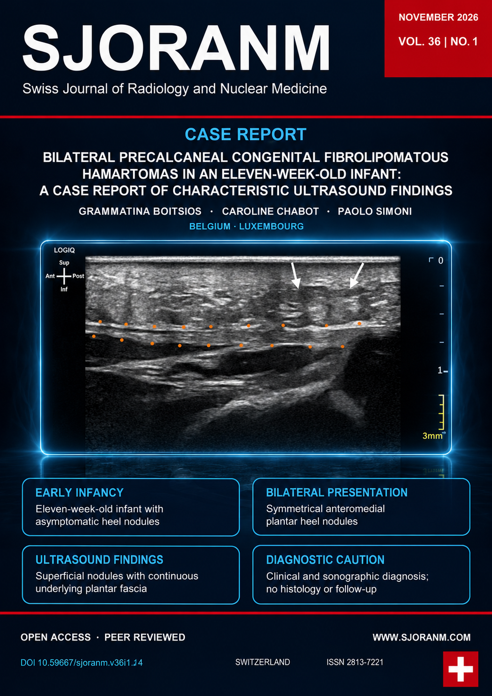
Precalcaneal congenital fibrolipomatous hamartomas (PCFHs) are benign subcutaneous nodules of infancy, typically located on the medial plantar heel and often bilateral. We report an eleven-week-old full-term male infant with symmetrical bilateral anteromedial plantar heel nodules. Ultrasound showed well-defined, oval, hypoechoic subcutaneous nodules. The plantar fascia remained continuous, with no sonographic evidence of involvement of the underlying muscles or osseous structures. Based on the age, characteristic location, bilateral symmetry, and sonographic features, bilateral PCFH was considered the most consistent diagnosis. No biopsy or MRI was performed, and no longitudinal follow-up was available. Conservative management with parental reassurance was recommended. Recognition of this characteristic clinical and sonographic pattern may help support conservative management and avoid unnecessary invasive investigation in typical cases.
References
1. Larralde de Luna M, Ruiz León J, Cabrera HN. [Pedal papules in newborn infants]. Med Cutan Ibero Lat Am. 1990;18(1):9-12. https://europepmc.org/article/MED/2214934
2. Fangman WL, Prose NS. Precalcaneal congenital fibrolipomatous hamartomas: report of occurrence in half brothers. Pediatr Dermatol. 2004;21(6):655-656. https://doi.org/10.1111/j.0736-8046.2004.21608.x DOI: https://doi.org/10.1111/j.0736-8046.2004.21608.x
3. Greenberg S, Krafchik BR. Infantile pedal papules. J Am Acad Dermatol. 2005;53(2):333-334. https://doi.org/10.1016/j.jaad.2005.02.035 DOI: https://doi.org/10.1016/j.jaad.2005.02.035
4. Ortega-Monzó C, Molina-Gallardo I, Monteagudo-Castro C, et al. Precalcaneal congenital fibrolipomatous hamartoma: a report of four cases. Pediatr Dermatol. 2000;17(6):429-431. https://doi.org/10.1046/j.1525-1470.2000.01816.x DOI: https://doi.org/10.1046/j.1525-1470.2000.01816.x
5. Tremaine AM, Paik AS, Metz B. Soft Subcutaneous Plantar Masses—Diagnosis. Arch Dermatol. 2012;148(9):1073-1078. https://doi.org/10.1001/archderm.148.9.1073-f DOI: https://doi.org/10.1001/archderm.148.9.1073-b
6. Laetsch Semadeni B, Mainetti C, Itin P, Lautenschlager S. Precalcaneal Congenital Fibrolipomatous Hamartomas: Report of 3 Additional Cases and Discussion of the Differential Diagnosis. Dermatology. 2009;218(3):260-264. https://doi.org/10.1159/000195175 DOI: https://doi.org/10.1159/000195175
7. Yang JH, Park OJ, Kim JE, Won CH, Chang SE, Lee MW, Choi JH, Moon KC. Precalcaneal congenital fibrolipomatous hamartoma. Ann Dermatol. 2011;23(1):92-94. https://doi.org/10.5021/ad.2011.23.1.92 DOI: https://doi.org/10.5021/ad.2011.23.1.92
8. Cho SB, Ha HI, Kang TW, Chung KY. Ultrasonographic assessment of piezogenic pedal papules. Clinical and Experimental Dermatology. 2009;34(7):e325-e326. https://doi.org/10.1111/j.1365-2230.2009.03278.x DOI: https://doi.org/10.1111/j.1365-2230.2009.03278.x
9. Choudhary AK, Adapa P. Precalcaneal congenital soft-tissue lesions in children: A case report of fibrous hamartoma of infancy and an approach to differential diagnosis. Radiol Case Rep. 2010;5(3):373. https://doi.org/10.2484/rcr.v5i3.373 DOI: https://doi.org/10.2484/rcr.v5i3.373
10. Saab ST, McClain CM, Coffin CM. Fibrous Hamartoma of Infancy: A Clinicopathologic Analysis of 60 Cases. American Journal of Surgical Pathology. 2014;38(3):394-401. https://doi.org/10.1097/PAS.0000000000000104 DOI: https://doi.org/10.1097/PAS.0000000000000104
11. Jacob CI, Kumm RC. Benign anteromedial plantar nodules of childhood: a distinct form of plantar fibromatosis. Pediatr Dermatol. 2000;17(6):472-474. https://doi.org/10.1046/j.1525-1470.2000.01825.x DOI: https://doi.org/10.1046/j.1525-1470.2000.01825.x
12. Kim HW, Yoo SY, Oh S, Jeon TY, Kim JH. Ultrasonography of Pediatric Superficial Soft Tissue Tumors and Tumor-Like Lesions. Korean J Radiol. 2020;21(3):341-355. https://doi.org/10.3348/kjr.2019.0343 DOI: https://doi.org/10.3348/kjr.2019.0343
13. Johnson CM, Navarro OM. Clinical and sonographic features of pediatric soft-tissue vascular anomalies part 2: vascular malformations. Pediatr Radiol. 2017;47(9):1196-1208. https://doi.org/10.1007/s00247-017-3906-x DOI: https://doi.org/10.1007/s00247-017-3906-x
Downloads
Published
Data Availability Statement
Not applicable. No datasets were generated or analyzed for this case report.
Issue
Section
License
Copyright (c) 2026 Grammatina Boitsios, Caroline Chabot, Paolo Simoni

This work is licensed under a Creative Commons Attribution 4.0 International License.
This license requires that reusers give credit to the creator. It allows reusers to distribute, remix, adapt, and build upon the material in any medium or format, even for commercial purposes.
 LinkedIn ↗
 X ↗
 VK ↗
 Truth Social ↗
 Bluesky ↗
 YouTube ↗