¹⁸F-FDG PET/CT in the Assessment of Suspected Mitral Prosthetic Valve Endocarditis
DOI:
https://doi.org/10.59667/sjoranm.v24i1.20Keywords:
FDG-PET/CT, Prosthetic valve endocarditis, Infective endocarditis, Mitral valve prosthesisAbstract
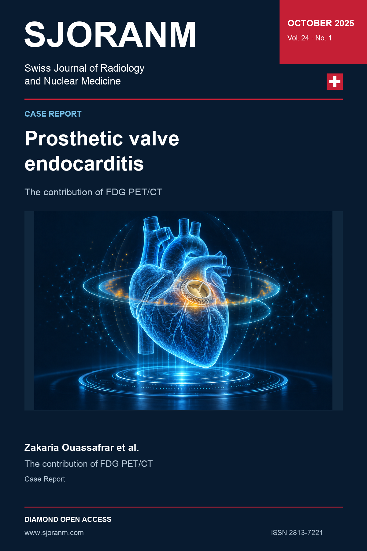
A 54-year-old woman with a mechanical mitral valve prosthesis presented with fever and tachycardia. Four pairs of blood cultures were positive for Escherichia coli. Echocardiography showed prosthetic obstruction and a mobile echogenic structure. The differential diagnosis included prosthetic valve endocarditis and prosthetic thrombosis. ¹⁸F-FDG PET/CT showed focal uptake in the prosthetic and periprosthetic region (SUVmax 2.6). Together with the clinical, microbiological and echocardiographic findings, this supported the diagnosis of prosthetic valve endocarditis. The case illustrates the contribution of PET/CT when echocardiography leaves the cause of prosthetic valve dysfunction uncertain.
References
1. Cahill TJ, Prendergast BD. Infective endocarditis. Lancet. 2016 ;387(10021):882-93. https://doi.org/10.1016/S0140-6736(15)00067-7 DOI: https://doi.org/10.1016/S0140-6736(15)00067-7
2. Bin Abdulhak AA, Baddour LM, Erwin PJ, Hoen B, Chu VH, Mensah GA, et al. Global and regional burden of infective endocarditis, 1990-2010: a systematic review of the literature. Glob Heart. 2014;9(1):131-43. https://doi.org/10.1016/j.gheart.2014.01.002 DOI: https://doi.org/10.1016/j.gheart.2014.01.002
3. Pant S, Patel NJ, Deshmukh A, Golwala H, Patel N, Badheka A, et al. Trends in infective endocarditis incidence, microbiology, and valve replacement in the United States from 2000 to 2011. J Am Coll Cardiol. 2015;65(19):2070-6. https://doi.org/10.1016/j.jacc.2015.03.518 DOI: https://doi.org/10.1016/j.jacc.2015.03.518
4. Murdoch DR, Corey GR, Hoen B, Miró JM, Fowler VG Jr, Bayer AS, et al. Clinical presentation, etiology, and outcome of infective endocarditis in the 21st century: the International Collaboration on Endocarditis-Prospective Cohort Study. Arch Intern Med. 2009;169(5):463-73. https://doi.org/10.1001/archinternmed.2008.603 DOI: https://doi.org/10.1001/archinternmed.2008.603
5. Gobbo MY, Meretta AH, Rosa D, Corneli M, Daquarti GJ, Masoli OH, et al. Tomografía por emisión de positrones en endocarditis infecciosa asociada a dispositivos y válvulas protésicas. Medicina (B Aires). 2020;80(1):17-22. https://pubmed.ncbi.nlm.nih.gov/32044737/
6. Fowler VG, Durack DT, Selton-Suty C, Athan E, Bayer AS, Chamis AL, et al. The 2023 Duke-International Society for Cardiovascular Infectious Diseases Criteria for Infective Endocarditis: Updating the Modified Duke Criteria. Clin Infect Dis. 2023 ;77(4):518-526. https://doi.org/10.1093/cid/ciad271 DOI: https://doi.org/10.1093/cid/ciad271
7. Vaidyanathan S, Patel CN, Scarsbrook AF, Chowdhury FU. FDG PET/CT in infection and inflammation–current and emerging clinical applications. Clin. Radiol. 2015, 70, 787–800. https://doi.org/10.1016/j.crad.2015.03.010 DOI: https://doi.org/10.1016/j.crad.2015.03.010
8. Chen W, Sajadi MM, Dilsizian V. Merits of FDG PET/CT and Functional Molecular Imaging Over Anatomic Imaging With Echocardiography and CT Angiography for the Diagnosis of Cardiac Device Infections. JACC Cardiovasc Imaging. 2018;11(11):1679–1691. https://doi.org/10.1016/j.jcmg.2018.08.026 DOI: https://doi.org/10.1016/j.jcmg.2018.08.026
9. Lancellotti P, Habib G, Oury C, Nchimi A. Positron Emission Tomography/Computed Tomography Imaging in Device Infective Endocarditis: Ready for Prime Time. Circulation. 2015;132(12):1076–1080. https://doi.org/10.1161/CIRCULATIONAHA.115.018521 DOI: https://doi.org/10.1161/CIRCULATIONAHA.115.018521
10. Aguadé Bruix S, Roque Pérez A, Cuéllar Calabria H, Pizzi MN. Cardiac 18F-FDG PET/CT procedure for the diagnosis of prosthetic endocarditis and intracardiac devices. Rev Esp Med Nucl Imagen Mol (Engl Ed). 2018;37(3):163-171. https://doi.org/10.1016/j.remnie.2018.02.001 DOI: https://doi.org/10.1016/j.remnie.2018.02.001
11. Delgado V, Ajmone Marsan N, de Waha S, Bonaros N, Brida M, Burri H et al. 2023 ESC Guidelines for the management of endocarditis. Eur Heart J. 2023 ;44(39):3948–4042. https://doi.org/10.1093/eurheartj/ehad193 DOI: https://doi.org/10.1093/eurheartj/ehad193
12. Erba PA, Sollini M, Conti U, Bandera F, Tascini C, De Tommasi SM, et al. Radiolabeled WBC scintigraphy in the diagnostic workup of patients with suspected device-related infections. JACC Cardiovasc Imaging. 2013;6:1075–1086. https://doi.org/10.1016/j.jcmg.2013.08.001 DOI: https://doi.org/10.1016/j.jcmg.2013.08.001
13. Erba PA, Conti U, Lazzeri E, Sollini M, Doria R, De Tommasi SM, et al. Added value of 99mTc-HMPAO-labeled leukocyte SPECT/CT in the characterization and management of patients with infectious endocarditis. J Nucl Med. 2012;53:1235–1243. https://doi.org/10.2967/jnumed.111.099424 DOI: https://doi.org/10.2967/jnumed.111.099424
14. Gotthardt M, Bleeker-Rovers CP, Boerman OC, Oyen WJ. Imaging of inflammation by PET, conventional scintigraphy, and other imaging techniques. J Nucl Med. 2010;51:1937–1949. https://doi.org/10.2967/jnumed.110.076232 DOI: https://doi.org/10.2967/jnumed.110.076232
15. Gomes A, Glaudemans AWJM, Touw DJ, van Melle JP, Willems TP, Maass AH, et al. Diagnostic value of imaging in infective endocarditis: a systematic review. Lancet Infect Dis. 2017 Jan;17(1):e1-e14. https://doi.org/10.1016/S1473-3099(16)30141-4 DOI: https://doi.org/10.1016/S1473-3099(16)30141-4
Downloads
Published
Issue
Section
License
Copyright (c) 2025 Zakaria Ouassafrar, Abdel Amide Gbadamassi, Halima Batani, Hafsa Bensimimoun, Amal Guensi

This work is licensed under a Creative Commons Attribution 4.0 International License.
This license requires that reusers give credit to the creator. It allows reusers to distribute, remix, adapt, and build upon the material in any medium or format, even for commercial purposes.
 LinkedIn ↗
 X ↗
 VK ↗
 Truth Social ↗
 Bluesky ↗
 YouTube ↗