Archives - Page 2
-
Vol. 09 No. 1 (2024)
Vol. 9 No. 1 (2024)The Intraosseous Schwannoma of the Upper Extremity Schwannomas are benign soft tissue tumors of neural origin, predominantly occurring in the head and neck regions due to their rich innervation. Intraosseous schwannoma (IOS) is an exceedingly rare form of schwannoma. This study aims to enhance the understanding of intraosseous schwannoma by reviewing cases affecting the upper extremity bones and providing a detailed analysis of their radiographic and magnetic resonance imaging (MRI) characteristics along with a review of the literature on these rare tumors. ---------------------------------------- The Inhomogeneous Focal Fat Distribution as Pseudolesions in a Patient with Pancreatic Carcinoma This case highlights the challenge of differentiating between hepatic pseudolesions caused by inhomogeneous fat distribution and true metastatic lesions. T1w opposed-phase imaging, diffusion-weighted imaging and hepatobiliary phase imaging after the administration of hepatocyte-specific contrast agents are crucial in making this distinction (4-5). The use of CEUS as a follow-up tool further aids in confirming the non-metastatic nature of such findings. ---------------------------------------- The Magnetic Resonance Imaging of a Rare Case of Primary Choriocarcinoma of the VulvaPrimary extrauterine choriocarcinoma is a rare condition, commonly reported in the cervix. Primary vulvovaginal choriocarcinoma has rarely been reported and review of literature reveals no case with MRI features for choriocarcinoma involving only the vulva. We report a rare case of primary choriocarcinoma of the vulva in a young woman, highlighting its Magnetic Resonance Imaging (MRI) features.
A 26-year-old female patient presented with complaints of menorrhagia, elevated serum Beta human chorionic gonadotropin (β-hCG) levels and a history of suction evacuation for partial molar pregnancy 4 months ago. Magnetic Resonance Imaging (MRI) of the pelvis revealed a normal uterus and adnexa. However, there was a well-defined T1 hypointense, T2 heterogenously hyperintense sublabial mass lesion with internal flow voids, few peripheral areas of restricted diffusion and few internal areas of T1 hyperintensity that bloomed on gradient images. On contrast administration, there was intense peripheral enhancement of the lesion, especially in the regions of restricted diffusion and also in the center of the lesion, where flow voids were observed. Few confluent non enhancing areas of necrosis were also present. In view of the clinical presentation, elevated β-hCG and imaging findings, a diagnosis of choriocarcinoma of the vulva was made and chemotherapy (EMA-CO regimen) was initiated, following which β-hCG decreased progressively and the patient is currently asymptomatic and follow up MRI revealed no abnormal significant residual lesion.
Awareness of a labial lesion being due to extrauterine choriocarcinoma in patients with elevated β-hCG is crucial for effective management and recovery. Our case uniquely emphasizes pretreatment MRI features as well as post-treatment imaging follow-up.
--------------------------------------------------
The Utility of multimodality imaging in a curious case of crippled childThe aim of the article is to establish the importance of devising and adhering to an imaging protocol for musculoskeletal imaging by presenting a case of Dysplasia epiphysealis hemimelica also known as Trevor’s disease in a 13 year old child with atypical presentation which proved to be a diagnostic challenge.
-
Vol. 08 No. 1 (2024)
Vol. 8 No. 1 (2024)A Collateral after Splenic Vein ThrombosisA 46-year-old man presented to the emergency department because of relapsing abdominal pain in the left abdominal quadrants. At admission, he was in good general condition and his vital signs were normal. The only abnormal laboratory findings were a C-reactive protein of 30 mg/L and thrombocytes of 127 G/L. An abdominal computed tomography (CT) scan showed a thrombosis of the distal splenic vein (panel A, double arrow) with the presence of a superior and an inferior spleno-mesenteric collateral (panel A, asterisks). The axial CT scan (panel V, arrows) and Doppler sonography (panel C) showed that the thick collaterals developed below the abdominal wall. A gastroscopy showed stage I fundic varices, but no esophageal varices. (1-10) This patient was diagnosed with a smoldering multiple myeloma 5 years earlier with regular follow-up in the hemato-oncology department. No other pro-thrombotic conditions were found and in a follow-up CT scan 6 months later the findings were unchanged despite anticoagulation. Splenic vein thrombosis is frequently diagnosed in the chronic stage, when collaterals have already developed (11-18). In this case, the presence of two major spontaneous shunts between the splenic vein and the superior mesenteric vein is preventing complications (19-23) such as porto-systemic encephalopathy and variceal bleeding.
-------------------------------------------------
The Role of 99mTc-HMDP Bone Scintigraphy in the Management of Neurogenic Para- Osteo-Arthropathies (NAOP)Neurogenic para-osteo-arthropathies (NAOP) are characterized by ectopic juxta-articular peri- or para-osseous ossifications. Diagnosis of these conditions is often delayed. They are frequently associated with central neurological disorders and are rarely observed in cases of peripheral neuropathy. Bone scintigraphy is crucial for determining the maturity of the ossification, optimizing surgical interventions, and preventing recurrence.
----------------------------------------------------
A Bilateral Ovarian GoitreOvarian goitre is classified within the category of single-tissue teratomas and represents a rare subtype of ovarian tumor. The histological identification of malignant variants has historically been contentious and inadequately assessed, primarily due to the lack of standardized diagnostic criteria and the tumor's infrequency. We present a case involving bilateral metastatic struma ovarii initially managed with conservative surgery, involving cystectomy on one side and radical oophorectomy on the other, as it was initially deemed benign. However, malignant transformation occurring seven years later necessitated a subsequent surgical intervention and adjunctive iratherapy.
-
Vol. 07 No. 2 (2024)
Vol. 7 No. 2 (2024)The ChatGPT may free time needed by the interventional radiologist for administration / documentationInterventional Radiology was, is and will be always busy. Our patients benefit from well planned and skilled performances done by physicians, who are free from bureaucracy. Can AI help? Perhaps it can help, first of all within the field of paperwork and endless documentation...later on perhaps AI will perform pleura drainage or PICC-Line insertion by itself. Time will show.
-
Vol. 07 No. 1 (2024)
Vol. 7 No. 1 (2024)The Contribution of Bone Scintigraphy in benign bone pathologyThis work provides an overview of the effectiveness of bone scintigraphy and its role in diagnosing various benign conditions in the Nuclear Medicine Department of the University Hospital of Fez. It analyzes the criteria for selecting scintigraphy as a diagnostic tool and outlines the principles, key features, and protocols involved. These details support clinicians in confirming their diagnostic reasoning when evaluating musculoskeletal lesions or bone abnormalities, guiding their choice of imaging modalities. Fibrodysplasia ossificans progressiva (FOP) is an extremely uncommon autosomal dominant condition marked by abnormalities in the big toes and the gradual development of extra-skeletal bone in specific anatomical formations.
-------------------------------------------
The Fibrodysplasia Ossificans Progressiva and Bone ScintigraphyFibrodysplasia ossificans progressiva (FOP) is an exceptionally rare autosomal dominant condition characterized by malformations of the big toes and progressive extra-skeletal ossifications in distinct anatomical patterns. In FOP, heterotopic ossification (HO) occurs episodically, resulting from spontaneous flare-ups or trauma, leading to cumulative disability. Bone scintigraphy with 99mTc-MDP combined with SPECT-CT is instrumental in early detection, diagnosis, staging, and disease monitoring by highlighting HO sites.
-
Vol. 06 No. 1 (2024)
Vol. 6 No. 1 (2024)The Multiple MyelomaThe second most common hematologic malignancy is the clonal proliferation of neoplastic plasma cells within the bone marrow. There is the presence of monoclonal immunoglobulins in the serum and/or urine. This results in anemia, myelosuppression, bone destruction, and clinical consequences of para-proteinemia on kidney function and other organ systems. The disease manifests through the acronym CRAB (hypercalcemia, renal impairment, anemia, and bone lesions). Less frequent manifestations of multiple myeloma are of extramedullary localizations. Myeloma cells can become independent of the bone marrow microenvironment, circulate freely in the blood, and infiltrate organs. This results in a high-risk state characterized by increased proliferation, evasion of apoptosis, and treatment resistance. It can affect any area of tissue. Most commonly it involves the pleura, lymph nodes, chest wall, liver, skin/soft tissue, lungs, CNS, genitourinary system, breast and pancreas. In patients with confirmed multiple myeloma, the diagnosis of extramedullary involvement is typically established by the presence of pathological soft tissue masses using radiological methods such as computed tomography (CT) scan, positron emission tomography/CT (PET/CT), magnetic resonance imaging (MRI), or ultrasound, along with biopsy or physical examination. The molecular mechanisms underlying the development of extramedullary multiple myeloma (EMM) have not been fully defined. Various cytogenetic abnormalities are observed, and some studies have generated genomic sequencing profiles that distinguish EMM from classic multiple myeloma. While plasma cell leukemia (PCL) and central nervous system (CNS) EMM indicate a poor prognosis, outcomes for other manifestations can be highly heterogeneous. Sensitive imaging modalities including PET/CT and MRI (Fig.1) are integral components of diagnosis and response assessment. Patients with extramedullary multiple myeloma (EMM) have a clear survival disadvantage.
---------------------------------------------
A Complex retrograde transarterial access and embolization of a giant internal iliac artery aneurysm after occlusion failure of Micro vascular plug (MVP) embolization and preceding EVAR in the distal ectatic Aorta and bilateral iliac artery aneurysmsThe therapy of pelvic aneurysms is minimally invasive through interventional radiology using various devices and embolic agents: 1. By using a covered stent/prosthesis to maintain antegrade flow when a landing zone, i.e., a vascular segment angiographically present into which the stent can be placed to bridge the neck of the aneurysm, thereby interrupting aneurysm perfusion. 2. Through complete embolization of the aneurysm using embolic agents/coils. 3. As performed in our case, by covering the aneurysm (Img 14 A-C) with a covered stent /prosthesis, following prior embolization of the branching vessels using macroembolic agents.
In cases where there is insufficient occlusion of the aneurysm with residual perfusion, secondary em- bolization becomes considerably more complex due to the occlusion of the anatomically antegrade access and the altered hemodynamics around the aneurysm causing retrograde collateralization.
Through detailed analysis of CT angiography, even in static CT, retrograde circulation could be identified, allowing for a minimally invasive secondary approach to the aneurysm without the need for general anesthesia and/or surgery. By completely eliminating the aneurysm, the risk of rupture in the elderly patient was minimally invasively eliminated. The treatment of this patient exemplifies the possibilities that embolization with a wide array of embolic agents and catheters offers in a hemodynamically very complex situation. -
Vol. 05 No. 1 (2024)
Vol. 5 No. 1 (2024)The Piracy? What Piracy?In discussing piracy herein, the reference is not to historical maritime raiders bestowed with titles by the British Empire, such as Sir Francis Drake, who appropriated gold from the Spaniards, themselves having previously stolen it from indigenous peoples ("Indians"). The act of pilfering from thieves is presently regarded with skepticism and presents distinct ethical quandaries. Rather, my focus pertains to instances wherein scientific and medical publications/ideas have been unlawfully appropriated. One might assume that such occurrences are antiquated or, at the very least, obsolete in contemporary times. Regrettably, they persist. Wherever opportunities emerge, certain transgressions ensue, irrespective of their moral permissibility. The adage "opportunity makes a thief" holds true. Within this discourse, I will scrutinize and evaluate specific behaviors exhibited by authors affiliated with publishing entities and those commonly termed "pirates". Various measures exist to comprehensively safeguard intellectual property against misappropriation. These encompass strategies such as confidentiality, distrust, heightened security awareness, precautionary measures, exercise of prudence, and the utilization of pre-print servers, among others.
------------------
The Regulatory Needs for Radiation Protection Devices based upon Artificial IntelligenceArtificial intelligence (AI) is increasingly employed in radiation protection, encompassing both medical devices and software. These technologies are integrated with AI throughout their manufacturing and application processes. This article underscores the imperative for comprehensive regulation in the utilization of AI. Decisions regarding AI application should not solely rest with manufacturers, medical professionals, or patients. Instead, an overarching "neutral" authority must be engaged to regulate, review, and enforce adherence to established protocols. The authors contend that relying on "self-regulation" within the free market, absent clear guidelines, proves to be inadequately effective and leads to patient's radiation protection safety issues.
-
Vol. 04 No. 1 (2024)
Vol. 4 No. 1 (2024)A Comparison of CT and MR Imaging of Cystic Renal Masses by Using the Bosniak Classification SystemPurpose:
To compare the efficacy of computed tomography (CT) and magnetic resonance imaging (MRI) in evaluating cystic renal masses using the Bosniak classification system.
Materials and Methods:
Between January 2010 and December 2020, a total of 844 patients with suspected renal masses underwent total or partial nephrectomy at the Department of Urology, Heidelberg University Clinic. Among them, 123 patients presented with cystic renal masses. Out of this cohort, 15 patients underwent preoperative examinations using both MRI and CT within a 6-week timeframe. These examinations were retrospectively analyzed by two radiologists employing the Bosniak classification system. Each lesion was assessed based on CT images for lesion localization, volume, number, and thickness of septa/wall, calcification, presence of enhancing soft-tissue components, and lesion density. Pathologic correlation was available for all lesions.
Results:
Out of the 844 patients, 15 underwent both CT and MR imaging examinations (male-to-female ratio: 2.75, age range: 43-75 years; mean age: 60.5 years). Based on CT images, there were one category I, one category II, three category IIF, three category III, and seven category IV lesions. Findings based on CT and MR images were concordant in eleven (73.3%) renal lesions, while in 4 (26.7%) masses, differences were observed. In three (20%) cases, MR imaging revealed more septa/wall than CT, resulting in an upgrade of the classification at MR imaging. In one (6.7%) lesion, MR imaging showed no wall and/or septa thickness compared with CT, leading to a classification downgrade. MR imaging results led to a cyst classification upgrade in three masses (from category II to III, n=1; IIF to III, n=2) and downgrade in one lesion (from category IIF to II). Pathologically, 11 lesions (73.3%) were malignant (9 Cystic Clear cell carcinoma and 2 papillary renal cell carcinoma), while 4 lesions were renal cysts without malignancy.
Conclusion:
The majority of findings from both CT and MR imaging were similar in suspected cystic renal masses. MR imaging demonstrated additional septa/wall or septa/wall thickening, potentially upgrading lesions from borderline category IIF to III compared to CT. This distinction may contribute to improved case management.----------------------------------------
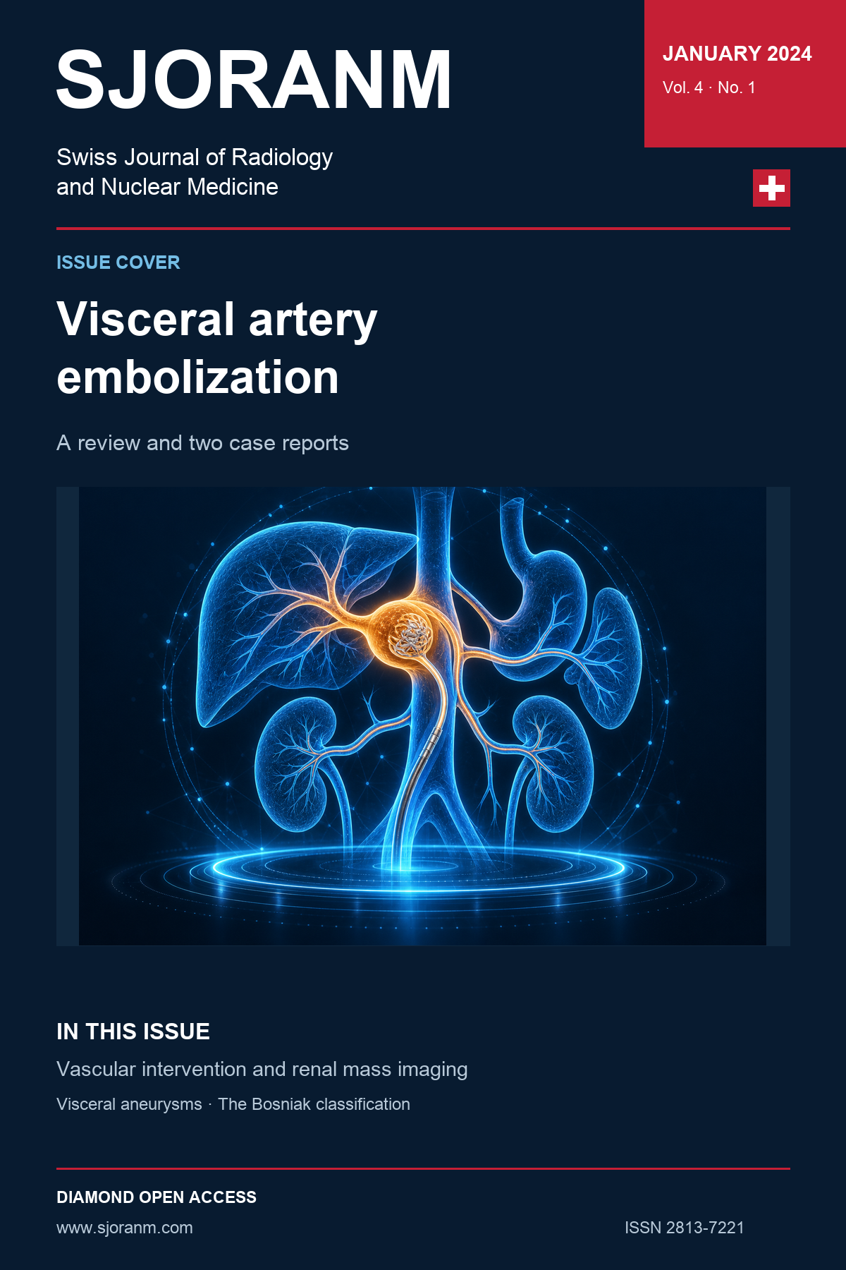
The Transarterial Embolization of Visceral Artery AneurysmsThe management of an aneurysm or pseudoaneurysm, including decisions regarding how, when, and with what method it should be treated, must always be determined through interdisciplinary consensus grounded in a thorough understanding of the existing literature. Consequently, today's preferred approach is embolization therapy or covered stenting. This method, performed in a minimally invasive manner, has been well-established due to its methodical low invasiveness and high effectiveness in achieving both technical and clinical success. It stands as a prominent choice among the available surgical and minimally invasive therapy options. The procedure, executed with advanced technology, utilizes the latest catheters and wires, coupled with expertise in various embolization methods. This comprehensive approach enables the treatment of acute bleeding in emergency cases, ensuring a nuanced and effective response.
-
Vol. 03 No. 1 (2023)
Vol. 3 No. 1 (2023)The Uncommon Cause of Recurrent PneumoniasA patient who presented with sudden shortness of breath, stridor, and recurrent laryngeal nerve paralysis was diagnosed with a tracheal diverticulum. The diagnosis was established through CT scans and confirmed via endoscopy. Distinctive CT findings revealed an air-filled, tubular structure located behind and slightly to the right lateral of the trachea, which communicated with it. Endoscopy demonstrated a narrow-mouthed diverticulum originating from the posterior right wall of the trachea, approximately 8 cm below the level of the true vocal cords. Successful medical treatment was administered, and the patient experienced an uncomplicated recovery.
------------------------------------------------
A Low Cellular Sclerosis of Anterior MediastinumConducting a CT-guided interventional procedure involving retrosternal pleural hydro-dissection of the juxta-aortic anterior mediastinum under local anesthesia revealed a rare histopathological condition known as benign mediastinal sclerosis. This challenging CT-guided intervention within the anterior mediastinum is scarcely documented in the existing scientific literature. In this presentation, we share our case along with relevant clinical data. What makes this case distinctive is that the clinicians initially questioned the representativeness of the biopsy sample we obtained. Consequently, an additional sample was acquired through a highly invasive video-assisted mediastinoscopy procedure under intubation anesthesia. Remarkably, this second sample yielded the same histopathological result.
Mediastinal sclerosis is an exceedingly rare condition, often referred to as the "burnt-out" stage of sclerosing mediastinitis. While infectious etiologies are frequently identified (Tab.1), the majority of cases remain idiopathic.
-
Vol. 02 No. 1 (2023)
Vol. 2 No. 1 (2023)The Early detection in cases of familial breast cancer predisposition: What is appropriate and beneficial for the individual seeking advice?A "high-risk situation" is present when the lifetime risk of developing breast cancer is ≥ 30%. Currently, the most accurate risk assessment is provided by the Tyrer-Cuzick model. This takes into account several factors including the presence of certain risk genes, age, family history of breast and ovarian cancer, as well as mammographic breast density. In addition to BRCA1 and BRCA2 several other risk genes are known that can be tested using gene panels. However specific familial risk constellations are prerequisites for indicating a genetic test. Prior to conducting a genetic test comprehensive counselling should take place and the individual seeking advice should be given time to consider. The individual seeking advice faces a series of questions regarding the potential implications of a genetic test which not only affect herself but also her environment.
In high-risk situations prophylactic mastectomy is an established surgical measure and intensified surveillance is a conservative approach. The latter includes semi-annual clinical breast examination with ultrasound starting from age 25 as well as an annual MRI mammography which exhibits the highest reliability compared to other imaging methods.
Medical consultation aims to enable a participatory decision-making process for the individual seeking advice. This requires comprehensive information for the individual seeking advice and incorporation of her preferences. Evidence-based decision aids from professional societies can improve the decisions of the individual seeking advice.
---------------------------------------------------------
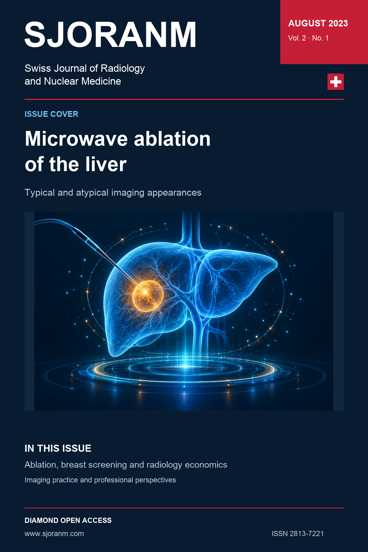
Typical and atypical appearance of microwave ablation zones in the liverLocal minimally invasive interventional procedures such as radiofrequency ablation (RFA) and microwave ablation (MWA) for the treatment of both primary hepatic malignancy and liver metastases of various origins have become increasingly popular in recent years. After treatment, is then the task of follow-up imaging with computed tomography (CT) and/or magnetic resonance imaging (MRI) to evaluate treatment success and to be aware of possible pitfalls that might give rise to misinterpretation.
---------------------------------------------------------
The Radiology in modern economics – business as usual?Until now the authors could identify three components to be critical to a well-functioning radiology system:
- The radiological self-perception and perception by others as a discipline.
- The radiologist as a human being.
- Depending on where the respective radiology department is located, the financial resources of radiology are very variable.
It is consensus in the medical community that radiologists play a key role in the diagnosis, treatment, and protection of patients [1]. In the past, various societal changes have led to a decline of reimbursement for radiological procedures and reductions in the quantity and quality of output. So-called demedicalization trends in radiology departments are taking place [2]. To make radiology fit for survival, several measures and strategy changes should be observed in the future. As we have learned from the various financial crises over the past 30 years, most western economies are driven primarily by profit maximization and ultra-rapid return on investment, leading to major inequalities in the distribution of wealth and health in society [3]. Will this be still acceptable in the future? Can radiology continue as it has been over the last decades? Business as usual? Will there be a significant financial deterioration in health system resources in the future, that will impact radiology negatively? To attempt to answer these questions, possible strategies for the survival of radiology as an independent discipline are discussed.
-
Vol. 01 No. 1 (2023)
Vol. 1 No. 1 (2023)Preface to the first issue of SJORANMhttps://doi.org/10.59667/sjoranm.v.1i.12 Radiology with its increasing numbers of steadily improving diagnostic and minimal invasive therapeutic methods in combination with the skills of diagnostic and therapeutic procedures in nuclear medicine are meanwhile essential tools on a very high level of patient treatment and care in tight cooperation with the clinicians. Our new Journal (SJORANM) wants to give the chance for quick online access to new ideas, new scientific research and results in the diagnostic and therapeutic field of Radiology and Nuclear Medicine.
-----------------------------------------------
Obituary Prof. Dr. med. Werner Wichmann, MDhttps://doi.org/10.59667/sjoranm.v.1i.13 Professor Dr. med. Werner Wichmann passed away on 26.01.2023
Werner Wichmann played a key role in shaping the emergence of the Swiss Society of Neuroradiology SGNR and the discipline itself. As an examiner and commission president, he was committed for many years to the standards to be met by a person working in clinical neuroradiology and at the same time was able to build trust in the next generation.
-----------------------------------------------
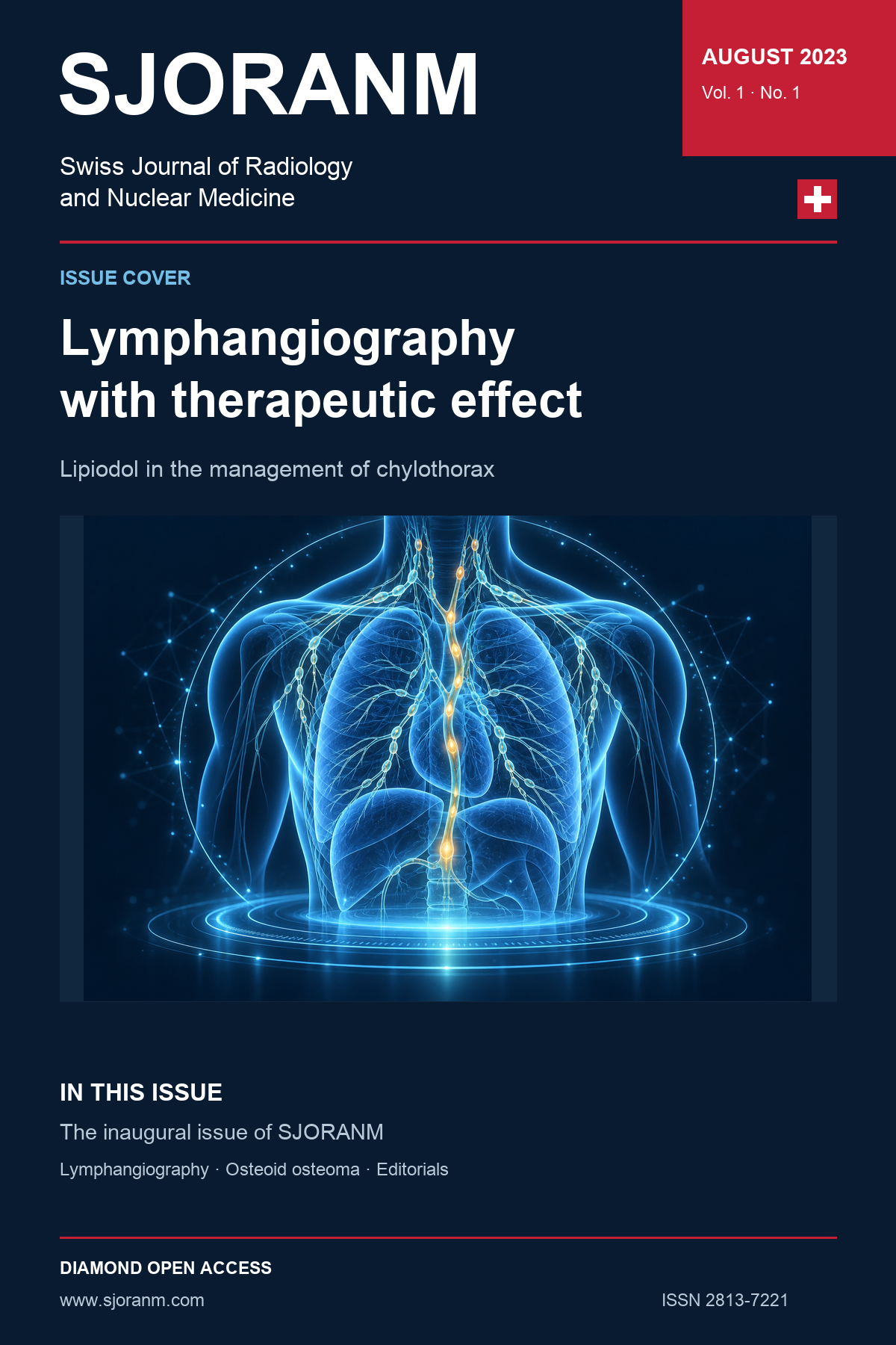
The Diagnostic lymphangiography with therapeutic effect: Since more than 70 years Lipiodol is used as an off-label standard of care for chylothorax - why not convert to on-label use?https://doi.org/10.59667/sjoranm.v1i.14 Purpose: The use of Lipiodol as a diagnostic agent and off-label therapeutic agent has been investigated in well over 395 publications listed in Pubmed under the key words: lymphangiography and chylothorax in the time period between 1921-2021.
While Lipiodol® ultra-fluid has been approved for use as a diagnostic agent in most countries, it can only be used off-label as a therapeutic agent for chylothorax, cholascos, and lymphatic leaks and fistulas. The therapeutic effects in chylothorax and the question why Lipiodol® ultra-fluid is still not approved for on-label use in the treatment of chylothorax are reviewed.
Background: Lipiodol was synthesized as iodized poppy seed oil by the French pharmacist Marcel Guerbet and first described by him in 1901. Over the past 12 decades, it has proven to be a reliable and versatile clinical theranostic agent. Lipiodol® ultra-fluid has been used (a) as a diagnostic contrast agent alone in the clarification and (b) so far only in off-label use as a therapeutic agent for chylothorax or cholascos, e.g. in cases of postoperative lymph leakages. Lipiodol® ultra-fluid has, to our knowledge, only very limited approvals as a therapeutic agent, in Switzerland only for transarterial chemo-embolization (TACE) for liver cancer. For decades and in numerous countries, Lipiodol® ultra-fluid has been in use as first-line treatment of chylothorax, cholascos, lymphatic leakage or lymphatic fistulae, avoiding additional interventions or surgery.
This review aims to assess in which countries Lipiodol® ultra-fluid is approved (a) as a primary diagnostic tool and (b) as a first line therapeutic agent. This review wants to reassess why the general therapeutic approval still lacks, and what could be done to achieve it.
-----------------------------------------------
Osteoid Osteoma – Synopsis of clinical, radiological, and therapeutical relevance of this rare entityhttps://doi.org/10.59667/sjoranm.v.1i.15 Characteristic of the osteoid osteoma is the so-called nidus enclosed in the tumor and produces a typical picture on X-ray. Since most physicians have little experience with the clinical picture of osteoid osteoma, this is an important reason for the often-long anamnesis time for osteoid osteoma (OO). The period of formation of OO includes the phase of strongest bone growth in childhood and adolescence. The most characteristic clinical symptom is nocturnal attacks of pain around the tumor, which occur independently of preceding physical activity and respond strikingly well to non-steroidal anti-inflammatory drugs (aspirin test). OO most frequently affects the long tubular bones of the lower extremities. Fifty percent of OO are found in the femur and tibia preferentially occurring in the corticalis of metaphysis and diaphysis. This is followed by the long tubular bones of the upper extremity and the short tubular bones. However, any other bone can also be affected. Differentially, osteoblastoma or osteosarcoma must be considered first and foremost. Most widely accepted therapy options are open surgery with en-bloc resection of the tumor, excochleation,minimally invasive percutaneous CT-guided radio or laser ablation. A conservative management by pharmaceutical pain therapy should be reserved for cases in which surgery must be refused either because of the precarious position of the nidus or because the patient's general condition does not permit it, or the patient does not consent to surgical interventions.