A Dual-time-point 18F-FDG PET/CT Imaging Unveiled Occult Hepatic Metastasis of Breast Cancer with Neuroendocrine Differentiation
A Case Report
DOI:
https://doi.org/10.59667/sjoranm.v32i2.14Keywords:
Delayed PET/CT, Breast cancer, occult metastasis, diagnostic imaging, liver metastasis, neuroendocrine differentiationAbstract
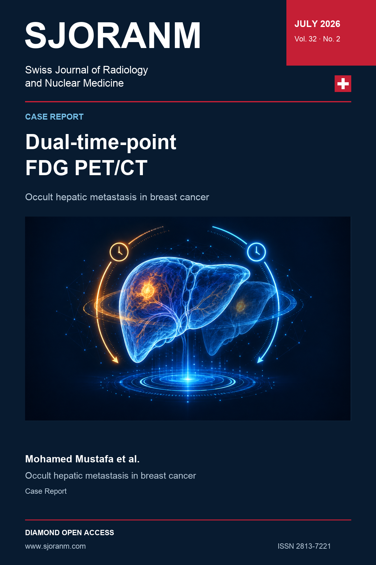
Breast cancer with neuroendocrine differentiation is a rare entity, often presenting diagnostic challenges due to its atypical imaging features. Standard imaging modalities may fail to detect early metastases, particularly when tumour markers rise in isolation. A 57-year-old woman with a history of right-sided invasive ductal carcinoma (estrogen receptor (ER) positive, progesterone receptor (PR) positive, human epidermal growth factor receptor 2 (HER2) negative) presented seven years after initial management with rising serum carcinoembryonic antigen (CEA) levels. Conventional imaging, including contrast-enhanced CT of the chest, abdomen and pelvis and standard-time 18F-FDG PET/CT, was negative. However, delayed PET/CT imaging (at 120 minutes) revealed a solitary metabolically active hepatic lesion with Maximum Standardized Uptake Value (SUVmax) 7.6, prompting further evaluation. Core biopsy identified the lesion as large cell neuroendocrine carcinoma (LCNEC) of breast origin, WHO grade 3. This case underscores the diagnostic value of delayed-phase 18F-FDG PET/CT in uncovering metabolically active occult liver metastasis from breast cancer, in those with biochemical recurrence and negative diagnostic imaging modalities including routine 18F-FDG PT/CT. Delayed imaging in dual time point 18F-FDG PET/CT detected a hepatic focal lesion guiding further lesion assessment by biopsy that properly characterized the lesion to have neuroendocrine differentiation, and directed further proper management achieving the concept of precision medicine.
References
1. Yang XR, Li YL, Li ZY, Chai XM. Primary hepatic neuroendocrine neoplasms: imaging characteristics and misdiagnosis analysis. Front Oncol. 2024 May 14;14. https://doi.org/10.3389/fonc.2024.1391663
2. Liu H, Shi J, Wilkerson ML, Lin F. Immunohistochemical Evaluation of GATA3 Expression in Tumors and Normal Tissues. American Journal of Clinical Pathology. 2012 Jul 1;138 [1]:57–64. https://doi.org/10.1309/AJCP5UAFMSA9ZQBZ
3. Al-Faham Z, Jolepalem P, Rydberg J, Wong CYO. Optimizing18 F-FDG Uptake Time Before Imaging Improves the Accuracy of PET/CT in Liver Lesions. J Nucl Med Technol. 2016 Jun;44 [2]:70–2. https://doi.org/10.2967/jnmt.115.169953
4. Lee JY, Song HS. Detection of Hepatic Metastasis from Early Delayed Images of Modified Dual-Time-Point 18F-FDG PET/CT Images in a Patient with Breast Cancer. Diagnostics. 2024 Jul 31;14 [15]:1653. https://doi.org/10.3390/diagnostics14151653
5. Annovazzi A, Rea S, Vici P, Fabi A, Sciuto R. Dual-time 18F-FDG PET/CT for the detection of liver metastases in breast cancer. Nuclear Medicine Communications. 2018 Dec;39 [12]:1183–9. https://doi.org/10.1097/MNM.0000000000000918
6. Kayani I, Bomanji JB, Groves A, Conway G, Gacinovic S, Win T, et al. Functional imaging of neuroendocrine tumors with combined PET/CT using68 Ga‐DOTATATE (DOTA‐ D Phe1 ,Tyr3 ‐octreotate) and18 F‐FDG. Cancer. 2008 Jun;112 [11]:2447–55. https://doi.org/10.1002/cncr.23469
7. Zhou J, Zhao R, Pan Y, Ju H, Huang X, Jiang Y, et al. The Diagnostic and Grading Accuracy of 68Ga-DOTATATE and 18F-FDG PET/MR for Pancreatic Neuroendocrine Neoplasms. Front Oncol. 2022 Feb 22;12:796391. https://doi.org/10.3389/fonc.2022.796391
8. Tirosh A, Kebebew E. The Utility of68 Ga-DOTATATE Positron-Emission Tomography/Computed Tomography in the Diagnosis, Management, Follow-Up and Prognosis of Neuroendocrine Tumors. Future Oncol. 2018 Jan 31;14 [2]:111–22. https://doi.org/10.2217/fon-2017-0393
9. Vegni F, De Stefano IS, Policardo F, Tralongo P, Feraco A, Carlino A, et al. Neuroendocrine neoplasms of the breast: a review of literature. Virchows Arch. 2024 Aug;485 [2]:197–212. https://doi.org/10.1007/s00428-024-03856-y
10. Rosen LE, Gattuso P. Neuroendocrine Tumors of the Breast. Archives of Pathology & Laboratory Medicine. 2017 Nov 1;141 [11]:1577–81. https://doi.org/10.5858/arpa.2016-0364-rs
11. Wang J, Wei B, Albarracin CT, Hu J, Abraham SC, Wu Y. Invasive neuroendocrine carcinoma of the breast: a population-based study from the surveillance, epidemiology and end results (SEER) database. BMC Cancer. 2014 Dec;14 [1]:147. https://doi.org/10.1186/1471-2407-14-147
12. Sun H, Dai S, Xu J, Liu L, Yu J, Sun T. Primary Neuroendocrine Tumor of the Breast: Current Understanding and Future Perspectives. Front Oncol. 2022 May 25;12:848485. https://doi.org/10.3389/fonc.2022.848485
Downloads
Published
Data Availability Statement
The data presented in this study are not publicly available due to institutional policies but are available on request from the corresponding author, MHM.
Issue
Section
License
Copyright (c) 2026 Dr. Mohamed Hossameldin Mustafa, Dr. Moumen Taha Katamesh, Dr. Yousry Wasif Nada, Dr. Batool Essa Albalooshi, Prof. Dr. Mohamad Mosaad ELLabban, MD, Dr. Mohamed Ali Elbeltagy, Prof. Dr. Sherin Yassin Wagih, Prof. Dr. Raafat Ali Saber, MD

This work is licensed under a Creative Commons Attribution 4.0 International License.
This license requires that reusers give credit to the creator. It allows reusers to distribute, remix, adapt, and build upon the material in any medium or format, even for commercial purposes.