Vol. 32 No. 2 (2026): Vol. 32 - No. 2
Egypt - https://doi.org/10.59667/sjoranm.v32i2.14
Mohamed Hossameldin Mustafa1*, Moumen Taha Katamesh1, Yousry Wasif Nada2, Batool Essa Albalooshi3, Mohamad Mosaad ELLabban4, Mohamed Ali Elbeltagy5, Sherin Yassin Wagih1, Raafat Ali Saber1
1Nuclear Medicine Department, Armed Forces College of Medicine, Cairo, Egypt
2Medical Oncology Department, Military Medical Academy, Cairo, Egypt
3Dubai Health, Dubai, United Arab Emirates
4Department of Anatomic Pathology, Faculty of Medicine, University of Port Said, Port Said, Egypt
5Pathology Department, Military Medical Academy, Cairo, Egypt
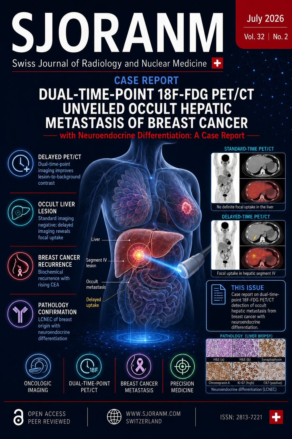
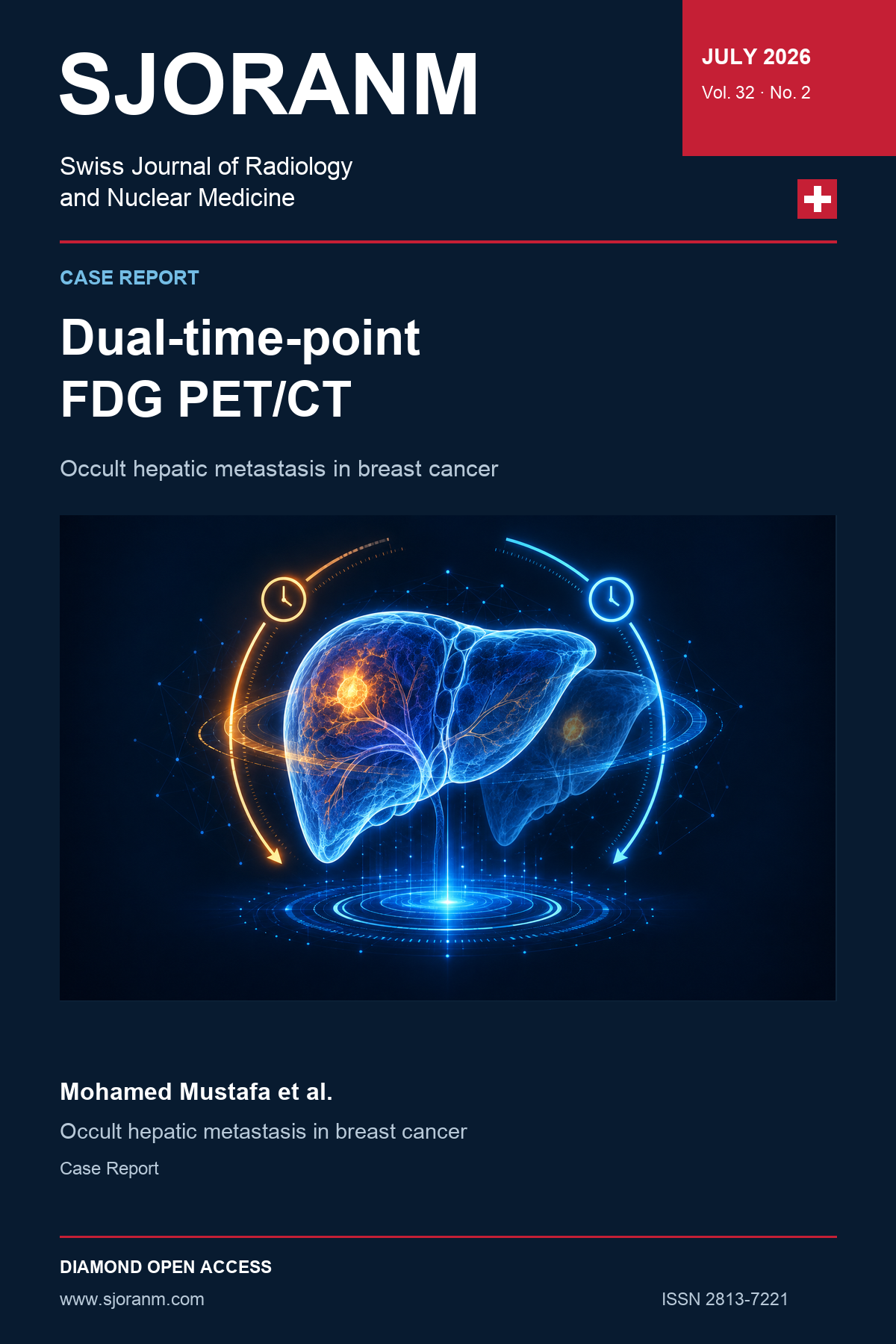
Breast cancer with neuroendocrine differentiation is rare and may be difficult to detect using standard imaging. A 57-year-old woman with previously treated ER- and PR-positive, HER2-negative invasive ductal carcinoma presented seven years later with rising carcinoembryonic antigen levels. Contrast-enhanced CT and standard-time ^18F-FDG PET/CT showed no evidence of recurrence. However, delayed PET/CT imaging at 120 minutes revealed a solitary hypermetabolic lesion in hepatic segment IV with an SUVmax of 7.6. Ultrasound-guided biopsy demonstrated a WHO grade 3 large-cell neuroendocrine carcinoma of breast origin. This case highlights the value of dual-time-point ^18F-FDG PET/CT in patients with biochemical evidence of recurrence but negative conventional imaging. Delayed imaging improved lesion-to-background contrast, enabled detection of an occult liver metastasis, guided biopsy, and supported appropriately tailored oncologic management.