Posterior Horn Dimensions of the Medial Meniscus and Predisposition to Posterior Horn Tears in Young Individuals: An MRI-Based Study
DOI:
https://doi.org/10.59667//sjoranm.v31i1.16Keywords:
Medial meniscus, posterior horn tear, size of posterior horn, dimensions of posterior horn, MRIAbstract
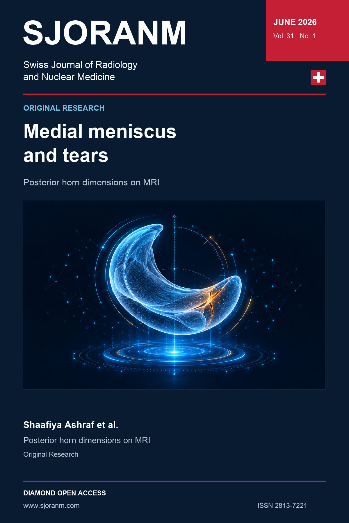
Objective: To evaluate the association between medial meniscus posterior horn dimensions and posterior horn tears in young individuals using MRI, and to determine an optimal cut-off value of posterior horn size associated with the presence of tears.
Material and Methods: This retrospective study included 554 patients aged 15–40 years, divided into tear and non-tear groups based on MRI findings. Meniscal size was assessed by measuring the anteroposterior (AP) diameter of the medial meniscus posterior horn on sagittal MRI images and the transverse diameter on coronal images. Measurements were compared between groups. Receiver operating characteristic (ROC) analysis was performed using the Youden Index to determine optimal cut-off values. Multivariable logistic regression analysis including posterior horn dimensions, age, sex, and body mass index (BMI) was performed to evaluate independent association with posterior horn tears. Interobserver reliability was assessed using intraclass correlation coefficient (ICC).
Results: The tear group demonstrated a significantly larger AP diameter of the medial meniscus posterior horn (18.77 ± 2.29 mm) compared with the non-tear group (14.36 ± 1.64 mm, p < 0.001). Similarly, the transverse diameter was significantly greater in the tear group (19.48 ± 2.75 mm) than in the non-tear group (14.45 ± 1.63 mm, p < 0.001). A cut-off value of ≥17 mm for AP diameter showed a sensitivity of 85.7%, specificity of 90.7%, and AUC of 0.943, while the same cut-off for transverse diameter demonstrated a sensitivity of 92.3%, specificity of 90.7%, and AUC of 0.955. Multivariable logistic regression confirmed that both AP and transverse diameters were independently associated with posterior horn tears after adjustment for age, sex, and BMI. Interobserver reliability showed excellent agreement for both AP (ICC = 0.91) and transverse (ICC = 0.92) measurements.
Conclusion: A significant association exists between increased size of posterior horn medial meniscus and its tears in young individuals, with tears occurring more frequently in those with a larger posterior horn.
References
1. Rath E, Richmond JC. The menisci: basic science and advances in treatment. British journal of sports medicine. 2000 Aug 1;34(4):252-7. https://doi.org/10.1136/bjsm.34.4.252 DOI: https://doi.org/10.1136/bjsm.34.4.252
2. Kurosawa H, Fukubayashi T, Nakajima H. Load-bearing mode of the knee joint: physical behavior of the knee joint with or without menisci. Clinical Orthopaedics and Related Research (1976-2007). 1980 Jun 1;149:283-90. DOI: https://doi.org/10.1097/00003086-198006000-00039
3. BB S. Transmission of the load in the knee joint with special reference to the role of the menisci II: Experimental results, discussion, and conclusions. Med. Eng.. 1979;8:220-8. DOI: https://doi.org/10.1243/EMED_JOUR_1979_008_051_02
4. Snoeker BA, Bakker EW, Kegel CA, Lucas C. Risk factors for meniscal tears: a systematic review including meta-analysis. journal of orthopaedic & sports physical therapy. 2013 Jun;43(6):352-67. https://doi.org/10.2519/jospt.2013.4295
5. Nguyen JC, De Smet AA, Graf BK, Rosas HG. MR imaging–based diagnosis and classification of meniscal tears. Radiographics. 2014 Jul;34(4):981-99. https://doi.org/10.1148/rg.344125202 DOI: https://doi.org/10.1148/rg.344125202
6. Pena E, Calvo B, Martinez MA, Palanca D, Doblaré M. Finite element analysis of the effect of meniscal tears and meniscectomies on human knee biomechanics. Clinical biomechanics. 2005 Jun 1;20(5):498-507. https://doi.org/10.1016/j.clinbiomech.2005.01.009 DOI: https://doi.org/10.1016/j.clinbiomech.2005.01.009
7. Jarraya M, Roemer FW, Englund M, Crema MD, Gale HI, Hayashi D, Katz JN, Guermazi A. Meniscus morphology: does tear type matter? A narrative review with focus on relevance for osteoarthritis research. InSeminars in arthritis and rheumatism 2017 Apr 1 (Vol. 46, No. 5, pp. 552-561). WB Saunders. https://doi.org/10.1016/j.semarthrit.2016.09.003 DOI: https://doi.org/10.1016/j.semarthrit.2016.11.005
8. Yokoi N, Bron AJ, Tiffany JM, Maruyama K, Komuro A, Kinoshita S. Relationship between tear volume and tear meniscus curvature. Archives of Othopedics. 2004 Sep 1;122(9):1265-9. https://doi.org/10.1001/archopht.122.9.1265 DOI: https://doi.org/10.1001/archopht.122.9.1265
9. Stone KR, Freyer A, Turek T, Walgenbach AW, Wadhwa S, Crues J. Meniscal sizing based on gender, height, and weight. Arthroscopy: The Journal of Arthroscopic & Related Surgery. 2007 May 1;23(5):503-8. https://doi.org/10.1016/j.arthro.2007.01.008 DOI: https://doi.org/10.1016/j.arthro.2006.12.025
10. Snoeker BA, Bakker EW, Kegel CA, Lucas C. Risk factors for meniscal tears: a systematic review including meta-analysis. journal of orthopaedic & sports physical therapy. 2013 Jun;43(6):352-67. https://doi.org/10.2519/jospt.2013.4295 DOI: https://doi.org/10.2519/jospt.2013.4295
11. Masquijo JJ, Bernocco F, Porta J. Discoid meniscus in children and adolescents: correlation between morphology and meniscal tears. Revista Española de Cirugía Ortopédica y Traumatología (English Edition). 2019 Jan 1;63(1):24-8. https://doi.org/10.1016/j.recot.2018.06.004 DOI: https://doi.org/10.1016/j.recote.2018.12.001
Downloads
Published
Data Availability Statement
Data is available with first three authors.
Issue
Section
License
Copyright (c) 2026 Shaafiya Ashraf, Arshed Hussain Parry, Sheetal Sharma, Arshad Bashir, Adnan Zahoor, Anil Sharma

This work is licensed under a Creative Commons Attribution 4.0 International License.
This license requires that reusers give credit to the creator. It allows reusers to distribute, remix, adapt, and build upon the material in any medium or format, even for commercial purposes.
 LinkedIn ↗
 X ↗
X ↗ VK ↗
VK ↗ Truth Social ↗
 Bluesky ↗
 YouTube ↗