Five Years of Silent Growth – An Anterior Abdominal Wall Spindle-Cell Neoplasm Suggestive of Synovial Sarcoma: A Case Report
DOI:
https://doi.org/10.59667/sjoranm.v36i4.14Keywords:
Synovial sarcoma, spindle-cell neoplasm, computed tomography, immunohistochemistry, Abdominal wall neoplasms, Soft-tissue sarcomaAbstract
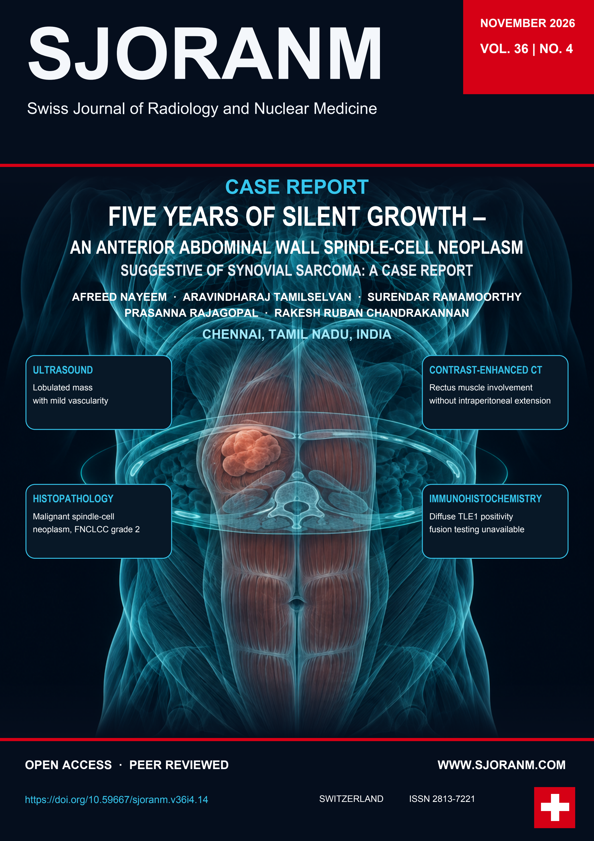
We report a 53-year-old man with a slowly enlarging, painless midline epigastric swelling of approximately five years’ duration. Ultrasonography showed a lobulated, heterogeneously hypoechoic lesion with mild internal vascularity. Contrast-enhanced CT showed a heterogeneously enhancing mass involving both rectus abdominis muscles, predominantly on the right, with no definite intraperitoneal extension. Thoracic staging showed no metastases. Wide local excision and mesh reconstruction were performed. Histopathology showed a malignant spindle-cell neoplasm, reported as FNCLCC grade 2, with predominantly fascicular and focal herringbone architecture. Immunohistochemistry showed diffuse TLE1 positivity and positivity for CD117 and S100. The findings were suggestive of synovial sarcoma. Molecular confirmation was unavailable. All microscopic margins were negative, with the closest measuring 1 mm. The patient received six cycles of ifosfamide followed by postoperative radiotherapy (50 Gy in 25 fractions). At clinical review five days after radiotherapy, he reported no new complaints. Post-treatment surveillance imaging had not yet been performed. This case illustrates the limitations of imaging and immunohistochemistry in establishing a definitive diagnosis without molecular confirmation.
References
1. McNeill J, Nguyen YV: Synovial sarcoma of the abdominal wall. Radiol Case Rep. 2007, 2:108. https://doi.org/10.2484/rcr.v2i4.108
2. Karadag O, Altundag K, Elkiran ET, Dikbas O, Gedikoglu G, Kars A: Anterior abdominal wall synovial sarcoma: a rare presentation. Am J Clin Oncol. 2005, 28:323-324. https://doi.org/10.1097/01.coc.0000144265.56369.a2
3. Jayaraman S, Rao SD, Govindarajan M. Synovial sarcoma of anterior abdominal wall. Indian J Surg. 2010;72(Suppl 1):293–295. https://doi.org/10.1007/s12262-010-0094-4
4. Alsaif HS. Primary synovial sarcoma of the abdominal wall: a case report and review of the literature. J Fam Community Med. 2008;15(3):123–125. https://pubmed.ncbi.nlm.nih.gov/23012178/
5. Bashir U, Moskovic E, Strauss D, Hayes A, Thway K, Pope R, Messiou C: Soft-tissue masses in the abdominal wall. Clin Radiol. 2014;69(10):e422–e431. https://doi.org/10.1016/j.crad.2014.06.006
6. Trigui R, Debaibi R, Guermazi A, Souai F, Dhaoui A, Chouchen A: Synovial sarcoma of the abdominal wall: a case report for a rare entity with a challenging treatment. Int J Surg Case Rep. 2023, 109:108596. https://doi.org/10.1016/j.ijscr.2023.108596
7. Khaladkar SM, Waghmode AM: Synovial sarcoma of anterior abdominal wall. Indian J Med Paediatr Oncol. 2020, 41:269-272. https://doi.org/10.4103/ijmpo.ijmpo_143_18
8. Murphey MD, Gibson MS, Jennings BT, Crespo-Rodríguez AM, Fanburg-Smith J, Gajewski DA: Imaging of synovial sarcoma with radiologic-pathologic correlation. Radiographics. 2006, 26:1543-1565. https://doi.org/10.1148/rg.265065084
9. de Haas RJ, Bonenkamp JJ, Flucke UE, de Rooy JWJ: Synovial sarcoma of the abdominal wall: imaging findings and review of the literature. J Radiol Case Rep. 2015, 9:24-30. https://doi.org/10.3941/jrcr.v9i2.1992
10. Dady M, Laamri I, El Allami Y, Ouhamou Y, Lakhdar S, El Hjouji A, Ait Ali A: Unusual location of synovial sarcoma of the abdominal wall: about a case and review of the literature. Asian J Res Surg. 2024;7(2):144–149. https://www.journalajrs.com/index.php/AJRS/article/view/204
11. Katyal A, Patel Y: Rare case of synovial sarcoma of anterior abdominal wall in a young Indian male. Int Surg J. 2017, 4:2358-2362. https://doi.org/10.18203/2349-2902.isj20172798
12. Virmani V, Sethi V, Fasih N, Ryan J, Kielar A: The abdominal wall lumps and bumps: cross-sectional imaging spectrum. Can Assoc Radiol J. 2014, 65:9-18. https://doi.org/10.1016/j.carj.2012.02.001
13. Mistarihi D, Obaideen A, Mukherji D, Faraj W: Incidental intra-abdominal synovial sarcoma. BMJ Case Rep. 2023, 16:e255628. https://doi.org/10.1136/bcr-2023-255628
14. Fertani Y, Bouhani M, Ben Naceur R, Jaidane O, Sahraoui G, Ziadi J, Ben Hassouna J, Ben Dhiab T. Synovial sarcoma of the abdominal wall with two-stage laced free latissimus dorsi reconstruction of a full-thickness defect: a case report and literature review. World J Surg Oncol. 2025, 23:327. https://doi.org/10.1186/s12957-025-03837-8
Downloads
Published
Data Availability Statement
The clinical and imaging findings relevant to this case are described in the article. No separate research dataset was generated.
Issue
Section
License
Copyright (c) 2026 Afreed Nayeem, Aravindharaj Tamilselvan, Surendar Ramamoorthy, Prasanna Rajagopal, Rakesh Ruban Chandrakannan

This work is licensed under a Creative Commons Attribution 4.0 International License.
This license requires that reusers give credit to the creator. It allows reusers to distribute, remix, adapt, and build upon the material in any medium or format, even for commercial purposes.
 LinkedIn ↗
 X ↗
 VK ↗
 Truth Social ↗
 Bluesky ↗
 YouTube ↗