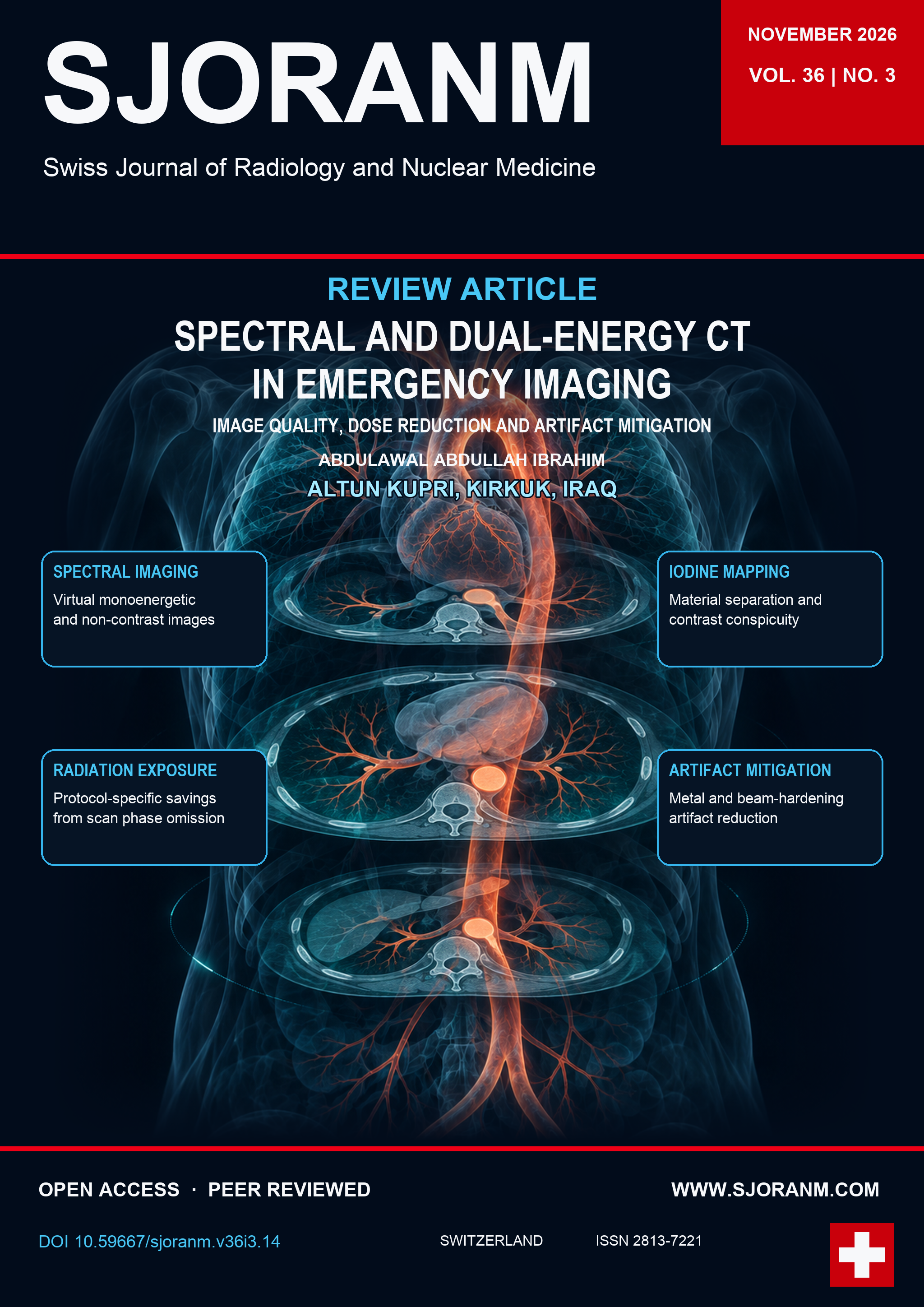
Spectral and Dual-Energy CT in Emergency Imaging: A Technical Review of Image Quality, Dose Reduction, and Artifact Mitigation
DOI:
https://doi.org/10.59667/sjoranm.v36i3.14Keywords:
Dual-energy CT, Spectral CT, Emergency imaging, Image quality, Radiation dose, Artifact reductionAbstract
Dual-energy and spectral computed tomography (DECT/spectral CT) extend conventional single-energy computed tomography (SECT) by measuring attenuation at two or more energy levels. This enables material decomposition, virtual monoenergetic imaging, and quantitative iodine mapping. In emergency radiology, these techniques may improve lesion visibility, reduce additional examinations and contrast medium requirements, and mitigate metal and beam-hardening artifacts when rapid and accurate diagnosis is required. This narrative review describes the underlying physical principles and available source-based, detector-based, and photon-counting technologies. It examines image quality, radiation dose, artifact reduction, and practical applications in emergency imaging.
References
1. Abdellatif W, Ebada MA, Alkanj S, Negida A, Murray N, Khosa F, Nicolaou S. Diagnostic accuracy of dual-energy CT in detection of acute pulmonary embolism: a systematic review and meta-analysis. Can Assoc Radiol J. 2021;72(2):285-292. https://doi.org/10.1177/0846537120902062 DOI: https://doi.org/10.1177/0846537120902062
2. Albrecht MH, Vogl TJ, Martin SS, et al. Review of clinical applications for virtual monoenergetic dual-energy CT. Radiology. 2019;293(2):260-271. https://doi.org/10.1148/radiol.2019182297 DOI: https://doi.org/10.1148/radiol.2019182297
3. Bamberg F, Dierks A, Nikolaou K, Reiser MF, Becker CR, Johnson TRC. Metal artifact reduction by dual energy computed tomography using monoenergetic extrapolation. Eur Radiol. 2011;21(7):1424-1429. https://doi.org/10.1007/s00330-011-2062-1 DOI: https://doi.org/10.1007/s00330-011-2062-1
4. Ehn S, Sellerer T, Muenzel D, et al. Assessment of quantification accuracy and image quality of a full-body dual-layer spectral CT system. J Appl Clin Med Phys. 2018;19(1):204-217. https://doi.org/10.1002/acm2.12243 DOI: https://doi.org/10.1002/acm2.12243
5. Forghani R, De Man B, Gupta R. Dual-energy computed tomography: physical principles, approaches to scanning, usage, and implementation: part 1. Neuroimaging Clin N Am. 2017;27(3):371-384. https://doi.org/10.1016/j.nic.2017.03.002 DOI: https://doi.org/10.1016/j.nic.2017.03.002
6. Gibney B, Redmond CE, Byrne D, Mathur S, Murray N. A review of the applications of dual-energy CT in acute neuroimaging. Can Assoc Radiol J. 2020;71(3):253-265. https://doi.org/10.1177/0846537120904347 DOI: https://doi.org/10.1177/0846537120904347
7. Graser A, Johnson TRC, Hecht EM, et al. Dual-energy CT in patients suspected of having renal masses: can virtual nonenhanced images replace true nonenhanced images? Radiology. 2009;252(2):433-440. https://doi.org/10.1148/radiol.2522080557 DOI: https://doi.org/10.1148/radiol.2522080557
8. Gupta R, Phan CM, Leidecker C, et al. Evaluation of dual-energy CT for differentiating intracerebral hemorrhage from iodinated contrast material staining. Radiology. 2010;257(1):205-211. https://doi.org/10.1148/radiol.10091806 DOI: https://doi.org/10.1148/radiol.10091806
9. Henzler T, Fink C, Schoenberg SO, Schoepf UJ. Dual-energy CT: radiation dose aspects. AJR Am J Roentgenol. 2012;199(5 Suppl):S16-S25. https://doi.org/10.2214/AJR.12.9210 DOI: https://doi.org/10.2214/AJR.12.9210
10. Im AL, Lee YH, Bang DH, et al. Dual energy CT in patients with acute abdomen; is it possible for virtual non-enhanced images to replace true non-enhanced images? Emerg Radiol. 2013;20:475-483. https://doi.org/10.1007/s10140-013-1141-9 DOI: https://doi.org/10.1007/s10140-013-1141-9
11. Johnson TRC. Dual-energy CT: general principles. AJR Am J Roentgenol. 2012;199(5 Suppl):S3-S8. https://doi.org/10.2214/AJR.12.9116 DOI: https://doi.org/10.2214/AJR.12.9116
12. Johnson TRC, Krauss B, Sedlmair M, et al. Material differentiation by dual energy CT: initial experience. Eur Radiol. 2007;17(6):1510-1517. https://doi.org/10.1007/s00330-006-0517-6 DOI: https://doi.org/10.1007/s00330-006-0517-6
13. Kalender WA, Perman WH, Vetter JR, Klotz E. Evaluation of a prototype dual-energy computed tomographic apparatus. I. Phantom studies. Med Phys. 1986;13(3):334-339. https://doi.org/10.1118/1.595958 DOI: https://doi.org/10.1118/1.595958
14. Lee YH, Park KK, Song HT, Kim S, Suh JS. Metal artefact reduction in gemstone spectral imaging dual-energy CT with and without metal artefact reduction software. Eur Radiol. 2012;22(6):1331-1340. https://doi.org/10.1007/s00330-011-2370-5 DOI: https://doi.org/10.1007/s00330-011-2370-5
15. Lell MM, Kachelrieß M. Recent and upcoming technological developments in computed tomography: high speed, low dose, deep learning, multienergy. Invest Radiol. 2020;55(1):8-19. https://doi.org/10.1097/RLI.0000000000000601 DOI: https://doi.org/10.1097/RLI.0000000000000601
16. McCollough CH, Leng S, Yu L, Fletcher JG. Dual- and multi-energy CT: principles, technical approaches, and clinical applications. Radiology. 2015;276(3):637-653. https://doi.org/10.1148/radiol.2015142631 DOI: https://doi.org/10.1148/radiol.2015142631
17. Mohammadinejad P, Kwapisz L, Fidler JL, et al. The utility of a dual-phase, dual-energy CT protocol in patients presenting with overt gastrointestinal bleeding. Acta Radiol Open. 2021;10:20584601211030658. https://doi.org/10.1177/20584601211030658 DOI: https://doi.org/10.1177/20584601211030658
18. Mohammed MF, Elbanna KY, Mohammed AME, et al. Practical applications of dual-energy computed tomography in the acute abdomen. Radiol Clin North Am. 2018;56(4):549-563. https://doi.org/10.1016/j.rcl.2018.03.004 DOI: https://doi.org/10.1016/j.rcl.2018.03.004
19. Monti CB, Zanardo M, Cozzi A, et al. Dual-energy CT performance in acute pulmonary embolism: a meta-analysis. Eur Radiol. 2021. https://doi.org/10.1007/s00330-020-07633-8 DOI: https://doi.org/10.1007/s00330-020-07633-8
20. Murray N, Darras KE, Walstra FE, Mohammed MF, McLaughlin PD, Nicolaou S. Dual-energy CT in evaluation of the acute abdomen. RadioGraphics. 2019;39(1):264-286. https://doi.org/10.1148/rg.2019180087 DOI: https://doi.org/10.1148/rg.2019180087
21. Nicolaou S, Yong-Hing CJ, Galea-Soler S, Hou DJ, Louis L, Munk P. Dual-energy CT as a potential new diagnostic tool in the management of gout in the acute setting. AJR Am J Roentgenol. 2010;194(4):1072-1078. https://doi.org/10.2214/AJR.09.2428 DOI: https://doi.org/10.2214/AJR.09.2428
22. Oberparleiter M, Vosshenrich J, Breit HC, Kaiser P, Hehenkamp P, Harder D, Boll DT, Zech CJ, Obmann MM. Dual-energy CT of acute bowel ischemia: influence on diagnostic accuracy and reader confidence. Eur Radiol. 2025;35(3):1405-1414. [cited as 2025a] https://doi.org/10.1007/s00330-024-11217-1 DOI: https://doi.org/10.1007/s00330-024-11217-1
23. Oberparleiter M, et al. Replacing true non-contrast imaging with DECT in GI bleeding demonstrates non-inferior diagnostic performance, reading time and confidence. Eur Radiol. 2025. [cited as 2025b] https://doi.org/10.1007/s00330-025-12191-y DOI: https://doi.org/10.1007/s00330-025-12191-y
24. Odedra D, Narayanasamy S, Sabongui S, Priya S, Krishna S, Sheikh A. Dual energy CT physics: a primer for the emergency radiologist. Front Radiol. 2022;2:820430. https://doi.org/10.3389/fradi.2022.820430 DOI: https://doi.org/10.3389/fradi.2022.820430
25. Pache G, Krauss B, Strohm P, et al. Dual-energy CT virtual noncalcium technique: detecting posttraumatic bone marrow lesions: feasibility study. Radiology. 2010;256(2):617-624. https://doi.org/10.1148/radiol.10091230 DOI: https://doi.org/10.1148/radiol.10091230
26. Patino M, Prochowski A, Agrawal MD, et al. Material separation using dual-energy CT: current and emerging applications. RadioGraphics. 2016;36(4):1087-1105. https://doi.org/10.1148/rg.2016150220 DOI: https://doi.org/10.1148/rg.2016150220
27. Pomerantz SR, Kamalian S, Zhang D, et al. Virtual monochromatic reconstruction of dual-energy unenhanced head CT at 65-75 keV maximizes image quality compared with conventional polychromatic CT. Radiology. 2013;266(1):318-325. https://doi.org/10.1148/radiol.12111604 DOI: https://doi.org/10.1148/radiol.12111604
28. Rajiah P, Parakh A, Kay F, Baruah D, Kambadakone AR, Leng S. Update on multienergy CT: physics, principles, and applications. RadioGraphics. 2020;40(5):1284-1308. https://doi.org/10.1148/rg.2020200038 DOI: https://doi.org/10.1148/rg.2020200038
29. Rassouli N, Etesami M, Dhanantwari A, Rajiah P. Detector-based spectral CT with a novel dual-layer technology: principles and applications. Insights Imaging. 2017;8(6):589-598. https://doi.org/10.1007/s13244-017-0571-4 DOI: https://doi.org/10.1007/s13244-017-0571-4
30. Reddy T, McLaughlin PD, Mallinson PI, et al. Detection of occult, undisplaced hip fractures with a dual-energy CT algorithm targeted to detection of bone marrow edema. Emerg Radiol. 2015;22(1):25-29. https://doi.org/10.1007/s10140-014-1249-6 DOI: https://doi.org/10.1007/s10140-014-1249-6
31. Si-Mohamed S, Dupuis N, Tatard-Leitman V, et al. Virtual versus true non-contrast dual-energy CT imaging for the diagnosis of aortic intramural hematoma. Eur Radiol. 2019;29(12):6762–6771. https://doi.org/10.1007/s00330-019-06322-5 DOI: https://doi.org/10.1007/s00330-019-06322-5
32. Sun H, Hou XY, Xue HD, et al. Dual-source dual-energy CT angiography with virtual non-enhanced images and iodine map for active gastrointestinal bleeding: image quality, radiation dose and diagnostic performance. Eur J Radiol. 2015;84(5):884-891. https://doi.org/10.1016/j.ejrad.2015.01.013 DOI: https://doi.org/10.1016/j.ejrad.2015.01.013
33. Wang TJ, Barrett S, Ali I, Khosa F, Nicolaou S, Murray N. Dual-energy CT in the acute setting: bowel trauma. Front Radiol. 2022;2:835834. https://doi.org/10.3389/fradi.2022.835834 DOI: https://doi.org/10.3389/fradi.2022.835834
34. Willemink MJ, Noël PB. The evolution of image reconstruction for CT: from filtered back projection to artificial intelligence. Eur Radiol. 2019;29(5):2185-2195. https://doi.org/10.1007/s00330-018-5810-7 DOI: https://doi.org/10.1007/s00330-018-5810-7
35. Willemink MJ, Persson M, Pourmorteza A, Pelc NJ, Fleischmann D. Photon-counting CT: technical principles and clinical prospects. Radiology. 2018;289(2):293-312. https://doi.org/10.1148/radiol.2018172656 DOI: https://doi.org/10.1148/radiol.2018172656
36. Wortman JR, Uyeda JW, Fulwadhva UP, Sodickson AD. Dual-energy CT for abdominal and pelvic trauma. RadioGraphics. 2018;38(2):586-602. https://doi.org/10.1148/rg.2018170058 DOI: https://doi.org/10.1148/rg.2018170058
Downloads
Published
Data Availability Statement
This narrative review is based on previously published literature cited in the reference list. No new datasets were generated for this review.
Issue
Section
License
Copyright (c) 2026 Abdulawal Abdullah Ibrahim

This work is licensed under a Creative Commons Attribution 4.0 International License.
This license requires that reusers give credit to the creator. It allows reusers to distribute, remix, adapt, and build upon the material in any medium or format, even for commercial purposes.
 LinkedIn ↗
 X ↗
 VK ↗
 Truth Social ↗
 Bluesky ↗
 YouTube ↗