
The Pediatric Ovarian Torsion at Two Years of Age
A Rare Gynecological Emergency
DOI:
https://doi.org/10.59667/sjoranm.v26i1.20Keywords:
Ovarian torsion, pediatric, young girl, normal ovaryAbstract
Introduction
Pediatric ovarian torsion is a relatively uncommon cause of acute abdominal pain. It carries significant risk of morbidity and possible mortality if not diagnosed and treated immediately.
Case presentation
In this report, we present a case of pediatric ovarian torsion in a two-year-old girl that was suspected on clinical assessment, diagnosed by ultrasound, and confirmed by exploratory laparoscopy. The right ovary was necrosed and oophorectomy was performed. This case report underlines the importance of a timely diagnosis to salvage the ovary.
Conclusion
This case report discusses the diagnosis of pediatric ovarian torsion including risk factors, symptoms, imaging modalities, and surgical diagnostics to improve diagnosis and shorten time to treatment. In addition, this case supports the use of laparoscopy for diagnosis of ovarian torsion if indicated by clinical suspicion and supplemental imaging studies.
--------------------------------------------------------------------------
Introduction
Ovarian torsion (OT) is a surgical emergency that can lead to impaired or lost fertility if the diagnosis and intervention are delayed. Complete or partial ovarian torsion occurs due to the twisting of the vascular pedicle of the ovary. The twisting of vascular pedicle causes obstruction of lymphatic and venous flow followed by obstructed arterial flow which if unrelieved ensues in ovarian infarction (1).
Pediatric ovarian torsion is an uncommon cause of acute abdominal pain in children. In adults, a large sized uterus provides support to the ovaries, which generally remain stably tucked, thus, reducing the chances of spontaneous torsion. Therefore, torsion in adults usually occurs when a cyst or mass develops within the ovary. Moreover, it is estimated to account for 3% of all cases of acute abdominal pain in adult women.
On the other hand, pediatric OT is an uncommon cause of acute abdominal pain. The small size of uterus provides space for ovaries to move freely at the cost of stability which predisposes ovaries to twist leading to torsion (2,3). It is pivotal to diagnose OT at the earliest, allowing a timely intervention to salvage the ovary as the delay in diagnosis can lead to impaired or lost fertility. Besides a missed or neglected OT can have serious complications such as intra-abdominal sepsis, septicemia, and death (4,5).
Case Presentation
A-2-year year old girl, presented to emergency department (ED) with right lower abdominal pain that has been persistent for 2 days. The pain was intermittent with no radiation. It was associated with anorexia, non-bilious vomiting, and constipation. No history of fever or diarrhea was forth-coming. There was no relevant past medical or surgical history. On examination, the child was lying comfortably in bed. The abdomen was soft and non-tender to palpation with normal bowel sounds on auscultation.
The child was afebrile with temperature of 37.2 °C. A cardiovascular system examination re-vealed sinus tachycardia with a pulse rate of 112 beats/minute, and blood pressure of 100/70 mmHg. Her respiratory rate was 26 breaths/minute, and her oxygen saturation was 100% on room air. Complete blood count revealed normal hemoglobin and platelet count with elevated white blood cell count (13.9 x10⁹/L). C-reactive protein concentration was 9 mg/l. Her urine analysis was normal. Kidney function test, liver function test, serum electrolytes, and other basic biochemical investigation results were within normal limits.
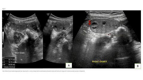
The patient was subjected to ultrasound examination of abdomen and pelvis, which showed an enlarged right ovary with multiple follicles and small peri-ovarian free fluid suggesting right ovarian torsion. The bowel loops were normal in caliber. No features of appendicitis or intussusception were seen.
Furthermore, evidence from clinical pictures and ultrasound findings lead to the decision to admit the patient and keep her fasting for exploratory laparoscopy and a possible right oophorectomy. The intraoperative laparoscopic findings demonstrated a torsed and completely gangrenous right ovary with no macroscopic viable ovarian tissue seen. The fallopian tube appeared healthy with intact fimbria. Minimal blood was seen in the right para-colic gutter and pelvis. Right oophorectomy was performed leaving behind the fimbria. The specimen was sent for histopathology examination which revealed widespread necrosis of the ovarian medulla and cortex, replaced by a dark brown tissue with hemorrhage and marked acute and chronic inflammation. No evidence of malignancy was seen. Post-operatively, the patient was discharged on pain medication and attached to pediatric surgery for follow-up.
Discussion
Adult females with OT usually present with typical clinical features which makes the diagnosis relatively easy. In comparison, the younger patients present with vague clinical features and are not able to articulate their symptoms clearly which makes the diagnosis challenging. A thorough clinical examination complimented by imaging in-vestigations is pivotal in clinching the diagnosis at the earliest to avert the deleterious consequences of OT.
OT is an important differential diagnosis in a female patient presenting with abdominal pain. The highest prevalence is seen in women of reproductive age group (2-5). It also constitutes as the fifth most common gynecological emergency in the reproductive age group. It has a cumulative prevalence of 2.7% and an incidence of 4.7 per 100,000 patients in women below 20 years of age (6-9). Pediatric and adolescent cases of OT account for about 15% of the total cases. Among the pediatric age group 52% cases of OT are reported between the age of 9 and 14 years. OT is uncommon in infants with only 6% of cases reported in children under 1 year old (10, 11). Right ovarian torsion is more common compared to the left side presumably due to presence of sigmoid colon on left side leaving little space for the ovary to twist (1). In the reproductive age an ovarian lesion (dermoid or functional cyst) predisposes to torsion as it makes the ovary heavier rendering it vulnerable to twist. However, in the pediatric and adolescent females usually no incriminating ovarian lesion is seen.
An earnest diagnosis of OT is essential to avoid any potential morbidity or mortality. Diagnosis of OT hinges primarily on clinical history and examination supplemented by imaging. However, confirmation of the diagnosis is usually made at the exploratory surgical procedure. Given the rarity of the condition and the non-specificity of the clinical and laboratory findings a timely diagnosis of OT can be challenging. Patients of OT usually present with nonspecific lower abdominal pain. The pain may be continuous or intermittent ranging in intensity from mild to severe. Associated symptoms like nausea and vomiting are commonly seen. A right sided OT can closely mimic appendicitis. Other differential diagnosis includes intussusception, gastroenteritis, and urinary tract infection.
The physical examination findings of fever, tachycardia and lower abdominal tenderness are nonspecific. Complete blood count, inflammatory markers and urinary analysis are commonly performed in suspected cases of OT to rule out other common mimics (6, 7). Aseptic pyuria is recognized in some patients (7). Ultrasound is the primary diagnostic tool for establishing the preoperative diagnosis. Ultrasound has a reported sensitivity of 92% and specificity of 96% in detecting OT. The ultrasound features of OT include an enlarged multifollicular ovary with free fluid around the ovary or in peritoneal cavity. Doppler ultrasound is a useful adjunct that increases the specificity. Doppler findings of reduced or absent vascular flow suggests diagnosis of OT. However, the preserved ovarian vascularity does not rule out torsion as ovary has a dual blood supply from ovarian and uterine artery. Additionally, an incomplete torsion may not compromise arterial blood flow to the ovary.
Detection of a twisted vascular pedicle on Doppler referred to as whirlpool sign constitutes a highly specific sign of torsion (11). In cases with non-diagnostic ultrasound findings additional studies like magnetic resonance imaging (MRI) and computed tomography (CT) are undertaken which can help in diagnosing as well as excluding the common differentials. However, in cases with ultrasound findings of OT treatment should be undertaken without subjecting the patient to CT or MRI. Surgical exploration is the definitive stan-dard for diagnosis and confirmation of OT via direct visualization.
Our patient presented with clinical features suspicious of OT which was further substantiated by ultrasound and finally confirmed by laparoscopy (Fig.2). Our patient recovered with no complications. Histology of the specimen demonstrated widespread necrosis of the ovarian medulla; cortex replaced by a dark brown hemorrhagic change. In a resource-constrained setting, it is a challenging to establish the diagnosis in a very limited time frame. Our patient had to undergo oophorectomy, but she did not have other complications. It is important to exclude OT with certainty in a young girl presenting with significant unexplained abdominal pain. Early diagnosis and treatment can salvage the ovary.
Conclusion
This case report highlights the importance of earnest timely diagnosis and emergent surgical intervention to salvage ovarian function and to limit any possible morbidity.
References
1. Sintim-Damoa A, Majmudar AS, Cohen HL, Parvey LS. Pediatric ovarian torsion: spectrum of imaging findings. Radiographics. 2017 Oct;37(6):1892-908. https://doi.org/10.1148/rg.2017170026
2. Rody A, Jackisch C, Klockenbusch W, Heinig J, Coenen-Worch V, Schneider HP. The conservative management of adnexal torsion—a case-report and review of the literature. European Journal of Obstetrics & Gynecology and Reproductive Biology. 2002 Feb 10;101(1):83-6. https://doi.org/10.1016/S0301-2115(01)00518-8
3. De Silva MH, Kolombage P, Kasthuri S. An ovarian torsion in a 2-year-old girl: a case report. Journal of Medical Case Reports. 2020 Dec;14(1):1-5. https://doi.org/10.1186/s13256-020-02518-2
4. Hasdemir PS, Eskicioglu F, Pekindil G, Kandiloglu AR, Guvenal T. Adnexal torsion with dystrophic calcifications in an adolescent: a chronic entity?. Case reports in obstetrics and gynecology. 2013 Dec 19;2013. https://doi.org/10.1155/2013/235459
5. Parelkar SV, Mundada D, Sanghvi BV, Joshi PB, Oak SN, Kapadnis SP, Shetty S, Athawale H, Multani P. Should the ovary always be conserved in torsion? A tertiary care institute experience. Journal of pediatric surgery. 2014 Mar 1;49(3):465-8. https://doi.org/10.1016/j.jpedsurg.2013.11.055
6. Yildiz A, Erginel B, Akin M, Karadağ CA, Sever N, Tanik C, Canmemiş A, Dokucu AI. A retrospective review of the adnexal outcome after detorsion in premenarchal girls. African Journal of Paediatric Surgery. 2014 Oct 1;11(4):304. https://doi.org/10.4103/0189-6725.143134
7. Servaes S, Zurakowski D, Laufer MR, Feins N, Chow JS. Sonographic findings of ovarian torsion in children. Pediatric radiology. 2007 May;37(5):446-51. https://doi.org/10.1007/s00247-007-0429-x
8. Poonai N, Poonai C, Lim R, Lynch T. Pediatric ovarian torsion: case series and review of the literature. Canadian Journal of Surgery. 2013 Apr;56(2):103. https://doi.org/10.1503/cjs.013311
9. De Silva MH, Kolombage P, Kasthuri S. An ovarian torsion in a 2-year-old girl: a case report. Journal of Medical Case Reports. 2020 Dec;14(1):1-5. https://doi.org/10.1186/s13256-020-02518-2
10. Swenson DW, Lourenco AP, Beaudoin FL, Grand DJ, Killelea AG, McGregor AJ. Ovarian torsion: Case–control study comparing the sensitivity and specificity of ultrasonography and computed tomography for diagnosis in the emergency department. European journal of radiology. 2014 Apr 1;83(4):733-8. https://doi.org/10.1016/j.ejrad.2014.01.001
11. Bhandari R, Khemani M, Mustafa A. Cases of management of paediatric tubo-ovarian torsion. International Journal of Reproduction, Contraception, Obstetrics and Gynecology. 2019 Jul 1;8(7):2888-95 https://doi.org/10.18203/2320-1770.ijrcog20193062
Downloads
Published
Data Availability Statement
The data are available from first two authors on reasonably request.
Issue
Section
License
Copyright (c) 2025 Mujahed Abdul Sattar Raheem, Arshed Hussain Parry, Hussam Hassan Ismail, Wael Hamed Ibrahim, Martin Corbally

This work is licensed under a Creative Commons Attribution 4.0 International License.
This license requires that reusers give credit to the creator. It allows reusers to distribute, remix, adapt, and build upon the material in any medium or format, even for commercial purposes.


