
A Comparison of diagnostic performance of CT with rectal contrast vs. CT with rectal and intravenous contrast for the diagnosis of acute appendicitis
DOI:
https://doi.org/10.59667/sjoranm.v26i1.18Keywords:
Acute appendicitis, Imaging, Radiation, iodine contrast, diagnostic performanceAbstract
Aim: Computed tomography (CT) is an essential investigation for the evaluation of suspected acute appendicitis (AA) owing to its high accuracy and the ability to provide an alternate diagnosis, however, there is still debate on the optimal CT technique with protocols varying between institutions. The present study aimed to compare the diagnostic accuracy of CT with rectal contrast (CT-RC) with that of CT with both rectal and intravenous contrast (CT-IVRC) in the diagnosis of AA.
Material and methods: CTs of 135 patients were analysed by 2 radiologists retrospectively. Clinical outcome was used as the final diagnosis. The diagnostic accuracy of each CT technique was calculated and compared with each other.
Results: There was strong agreement inter- and intra-observer agreement for the diagnosis of AA (kappa= 0.76, 0.87, 0.89 and 0.91 for CT-RC and CT-IVRC, respectively). The sensitivity, specificity, positive predictive value, negative predictive value and diagnostic accuracy of CT-RC and CT-IVRC for the diagnosis of AA were not statistically different from each other (p-value>0.05 for all comparisons). The accuracy of two CT protocols in diagnosing AA ranged from 82% to 88%. The area under the curves (AUC) for diagnosing AA on CT-RC and CT-IVRC for two observers were 0.87. 0.9, 0.89 and 0.91 respectively.
Conclusions: CT-RC proved to be as accurate as CT-IVRC in the diagnosis of AA. CT with rectal contrast alone could be performed in suspected cases of AA particularly in patients with contraindications to intravenous contrast administration.
--------------------------------------------------------------------
Introduction
Acute appendicitis (AA) constitutes one of the im-portant causes of abdominal pain and is a frequent reason for emergency department (ED) visits. AA is the most common cause of hospitalization in young patients presenting with acute abdomen (1). AA has an estimated annual occurrence of 5.7–50 per 100,000 population per year with a peak incidence between the age of 10 -30 years (1).
The clinical data and laboratory information may not be always sufficient to diagnose or exclude AA or to provide an alternative diagnosis. Although appendectomy has been the standard of care for the management of AA, recently there has been a gradual shift towards conservative management of AA (2-3). However, before a firm management decision is made it is essential to establish a confident diagnosis of AA. Imaging comprising of ultrasonography (US), computed tomography (CT), and magnetic resonance imaging (MRI) all can be used to clinch the diagnosis of AA (3). US is the preliminary diagnostic tool (4). However, abdominal CT has superseded US for the evaluation of suspected AA in most centres owing to its high accuracy and the ability to provide an alternate diagnosis in non-appendicitis patients (5). It has been observed that CT decreases the negative appendectomy rates without increasing the rate of AA-related complications like perforation or peritonitis (6). MRI is reserved for specific clinical scenarios where the US may be unable to provide a diagnosis and CT is contraindicated like in pregnancy (7, 8).
The debate for the selection of optimal CT technique in the evaluation of suspected AA is still unresolved and different centres opt for different protocols (5). CT with administration of both intravenous (IV) and positive oral contrast has been the traditional technique with arguably the highest diagnostic yield (5, 9). The widespread use of this technique was based on the premise that this is the standard body imaging technique capable of maximising the visualisation of the appendix and allowing for the identification of extra-appendiceal pathologies (5). Despite its remarkably high accuracy for diagnosing AA, this technique is not necessarily the optimal or safest technique. Administration of IV contrast is fraught with many potential risks including the risk of anaphylactic reactions, risk of contrast-induced nephropathy (CIN), potential risk of extravasation and increased cost (10). The use of positive oral contrast is associated with time delays as it requires the passage of contrast to the caecum (11). The alternative protocols used by different centres across the world include IV contrast alone, positive oral contrast alone, rectal contrast alone, both IV and rectal contrast and no use of contrast (12-17).
The use of positive oral contrast and rectal contrast can aid in the easy detection of the appendix when the caecum and terminal ileum are opacified with contrast and secondly, filling of the lumen of an appendix with contrast essentially rules out appendicitis (5). Positive oral contrast is associated with a time delay of approximately 1-2 hours which is the usual transit time and secondly, in 18-30% of patients the contrast fails to reach the caecum (5). Additionally, oral contrast will not be tolerated by patients with nausea or vomiting. Rectal contrast is associated with shorter delays of around 20 minutes and acceptable levels of patient discomfort (18). The use of rectal contrast-enhanced CT (CT-RC) has been studied in isolation and has been found useful with a sensitivity of 98% and a diagnostic accuracy of 98% (19). However, a direct comparison between CT-RC and rectal contrast plus IV contrast-enhanced CT (CT-IVRC) is lacking. The purpose of this study was to compare the diagnostic accuracy of CT-RC with that of CT-IVRC in the diagnosis of AA.
Methods
Study design and patient cohort
This was a single-centre retrospective cross-sectional analytical study conducted for a period of one year, between January 2020 to December 2020. The study was approved by the institutional research committee. Due to the retrospective nature of the study, the requirement for patient consent was waived.
One hundred thirty-five patients who presented to ED with abdominal pain with a clinical suspicion of AA and underwent abdominal CT with both rectal and IV contrast were retrospectively enrolled in the study. Abdominal CT was performed after administration of rectal contrast followed by a second scan after administration of IV contrast according to the departmental protocol. Patients who had only rectal or IV-enhanced scans were excluded. The final diagnosis of AA was confirmed by surgery/histopathology or conservative management with follow-up for more than 2 months.
We searched our electronic database for cases of non-traumatic acute right lower quadrant or lower abdominal pain who reported to ED and underwent abdominal CT with a clinical suspicion of AA in the year 2020. Radiological, clinical, surgical, pathological and follow-up data were collected and analysed retrospectively.
Among the entire cohort of 135 patients, 56 pa-tients received a final diagnosis of AA. Taking surgery or conservative management for AA as the reference standard we evaluated the diagnostic accuracy of CT-RC and compared it with that of combined CT-IVRC.
Data collection
CT acquisition protocol and image interpreta-tion
All CT scans were performed on 64-slice Multi-detector CT. All patients underwent a CT-RC first. Rectal contrast was administered in the left lateral position using a rectal tube and 1-1.5 litre of normal saline mixed with 20-30ml of iodinated contrast. From the initial left lateral position patients were rolled into a supine and then right lateral position to allow the passage of contrast into the caecum. After 20 minutes first scan was acquired in a single breath-hold after setting up the patient in a head-first supine position in the CT gantry. The following scanning parameters were used: slice thickness 1-1.5 mm, tube voltage 100-120 kVp, tube current of 90-130mAs and a beam pitch of 1.5. The tube current was regulated by an automatic exposure control sys-tem. Images were reconstructed using a reconstruction increment of 0.7mm into a slice thickness of 1 mm. Using the same position and parameters a second scan was obtained after IV contrast administration in the portal venous phase.
To avoid any bias the CT scans were anonymised by a third radiologist (B.A) who was not involved in reading the images and then presented to the interpreting radiologists. Each patient had two sets of images including CT-RC and CT-IVRC. The CT images were analysed independently by two radiologists, one consultant and another resident with 15 and 4 years of experience, respectively, who were blinded to the clinical and outcome data.
The CTs were analysed for the following charac-teristics:
- Visualization or non-visualization of an appendix
- Fluid-filled or air-filled appendiceal lumen
- maximum outer diameter of an appendix
- presence of fecolith in the lumen of appendix
- presence of peri-appendiceal fat stranding
- enlarged surrounding nodes
- presence of peri-appendiceal free fluid or collection
- presence of extra-luminal air.
Appendicitis was defined according to the established criteria of maximal outer diameter >6mm with surrounding inflammation like peri-appendiceal fat stranding or fluid with a non-opacified lumen (20).
Statistical analysis
Data were analyzed using the Statistical Package for the Social Sciences (SPSSInc. Chicago, IL, version 21.0). Continuous variables were expressed as means and standard deviations. Categorical variables were expressed as counts and percentages. Fisher’s exact test was used to examine the categorical variables. Two sample student t-tests was used for the comparison of continuous variables when the data was normally distributed, while the Mann-Whitney U test was used when the data was not normally distributed. A p-value less than 0.05 was considered statistically significant. Cohen’s kappa was used to determine inter-observer reliability for reading CT images.
Results
135 patients were enrolled into the study comprising 69 (51%) male and 66 (49%) female patients. The mean age of the study population was 32.26±11.73 years. Based on surgery/histo-pathology or conservative management 56 (41.5%) patients received a final diagnosis of AA and the remaining 79 (58.5%) were found to have an alternate diagnosis. Among the baseline characteristics only WBC counts and CRP levels were significantly higher in the AA group compared to the non-AA group (Table 1).
Computed tomography findings of acute appendicitis
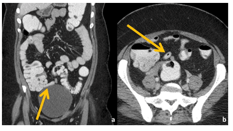
Various CT findings were correlated between the AA and non-AA groups. The appendix diameter was significantly higher in AA patients compared to non-AA patients (10 mm vs 5.2 mm) (p-value <0.001). Similarly, wall thickening, peri-appendiceal stranding, peri-appendiceal nodes, peri-appendiceal fluid, presence of appendicolith and wall interruption had a statistically significant association with a diagnosis of AA, whereas the presence of intraluminal contrast or intraluminal air and absence of abnormal wall enhancement were negative predictors of AA on both CT-RC and CT-IVRC (all p-values <0.05) (Table 2 and 3) (Figs. 1-5).
Agreement between the two observers for various parameters on CT-RC and CT-IVRC:
A consultant and a radiology resident were the interpreters. There was good agreement (k= 0.76) between the two readers in diagnosing AA on CT-RC (Table 2). However, there was almost perfect agreement (k= 0.87) between the two readers in diagnosing AA on CT-IVRC (Table 3).
For individual parameters, the level of agreement was moderate (kappa= 0.41 – 0.60) between the two observers in interpreting some parameters like wall thickening, intraluminal contrast, or intraluminal air, peri-appendiceal nodes and peri-appendiceal fluid and good to very good agreement (k > 0.60) in interpreting other parameters (peri-appendiceal stranding and appendicolith) on CT-RC. However, the level of agreement in-creased marginally between the two observers on CT-IVRC for various individual parameters (good agreement (k>0.60) for all the parameters except periappendiceal lymph nodes) (Table 3). Observer 1 was able to provide a diagnosis in all cases on both CT-RC and CT-IVRC, whereas Observer 2 was not able to provide any diagnosis in two cases on both CT-RC and CT-IVRC.
Intra-observer agreement for both readers:
The intra-observer agreement for various parameters for both observers is given in Table 4. The level of agreement was marginally better for both the observers on CT-IVRC.
Comparison of diagnostic performance of two CT protocols in detecting acute appendicitis:
The receiver operator characteristic (ROC) curve showed that the area under the curve (AUC) for diagnosing AA on CT-RC for two observers was 0.87 and 0.9, respectively. The AUC for diagnosing AA on CT-IVRC for two observers was 089 and 0.91, respectively (Table 5). The sensitivity, specificity, PPV, NPV and diagnostic accuracy of CT-RC were not significantly different between CT-IVRC and CT-RC for both observers (Table 5).
Discussion
CT is a sensitive and specific imaging tool for diagnosing AA, with pooled sensitivity and specificity of 96% and 92% (1). The use of CT in this clinical setting has been shown to decrease negative laparotomy rates and improve patient care (6). Despite its widespread use, there is still no firm consensus over the optimal CT technique for the diagnosis of AA. So, different institutions follow different CT protocols. CT with IV, oral contrast, rectal contrast and non-contrast CT are all employed for the diagnosis of AA (10). Non-contrast CT can be performed rapidly without the attendant risks and discomfort of contrast, but it may fail to detect AA in some cases especially when the reader is inexperienced.
IV contrast administration entails the risks of allergic reactions and CIN. CT with oral contrast is time-consuming, may lead to diagnostic delay and may not be feasible in patients with nausea and vomiting. CT-RC which can be performed rapidly is free from potential allergic reactions associated with IV contrast administration, and, therefore, may be the preferred initial technique in the diagnostic workup of suspected AA (12).
The present study demonstrated that the two CT protocols employing only CT-RC and CT-IVRC had comparable diagnostic accuracy for the diagnosis of AA. The AUC for both protocols was around 0.90 for both observers. The major advantage of the current study was that it provided a direct comparison between CT-RC and CT-IVRC. In contrast to previous studies (15-19), no difference was found in the ability (diagnostic accuracy, sensitivity, specificity, PPV, or NPV) to identify patients with AA. Similarly, they did not differ significantly in their ability to provide an alternative diagnosis in non-appendicitis patients.
CT-RC is equally accurate, although less sensitive, compared to combined CT with oral and IV contrast and significantly superior to non-contrast CT for the diagnosis of AA (15). S. Walker et. al in a study using CT-RC obtained a sensitivity of 94%, specificity of 100%, and accuracy of 96%, in diagnosing AA (16).
Mittal V.K et.al performed a randomized controlled trial to compare the accuracy of CT-RC alone with a triple contrast CT (oral, rectal, and IV contrast administration) and concluded that CT-RC had a comparable diagnostic performance and was better tolerated by patients, financially cheaper and reduced the time to diagnosis and negative appendectomy rate with no missed diagnosis (20).
Our results reiterate that CT-RC has several advantages. It is less time-consuming, tolerated well by patients with no potential hazards of IV or oral contrast and has a comparable diagnostic accuracy. Additionally, the diagnostic accuracy was almost similar for both experienced and relatively inexperienced radiologists.
However, retrospective design, single centre and small sample size are some important limitations of the study.
Conclusion
CT-RC has a comparable diagnostic performance compared to CT-IVRC in the detection of AA. CT-RC could be performed in suspected cases of AA particularly where there is a contraindication to IV contrast administration or if there is a risk of developing a severe adverse reaction.
References
1. Di Saverio S, Podda M, De Simone B, Ceresoli M, Augustin G, Gori A et al. Diagnosis and treatment of acute appendicitis: 2020 update of the WSES Jerusalem guidelines. World J Emerg Surg 2020;15:1-42. https://doi.org/10.1186/s13017-020-00306-3
2. CODA Collaborative. A randomized trial comparing antibiotics with appendectomy for appendicitis. N Engl J Med 2020;383(20):1907-19. https://doi.org/10.1056/NEJMoa2014320
3. Talan DA, Saltzman DJ, Mower WR, Krishnadasan A, Jude CM, Amii R et al. Antibiotics-first versus surgery for appendicitis: a US pilot randomized controlled trial allowing outpatient antibiotic management. Ann Emerg Med 2017;70(1):1-11.e9. https://doi.org/10.1016/j.annemergmed.2016.08.446
4. Pinto F, Pinto A, Russo A, Coppolino F, Bracale R, Fonio P et al. Accuracy of ultrasonography in the diagnosis of acute appendicitis in adult patients: review of the literature. Crit Ultrasound J 2013;5 Suppl 1:S2. https://doi.org/10.1186/2036-7902-5-S1-S2
5. Paulson EK, Coursey CA. CT protocols for acute appendicitis: time for change. AJR Am J Roentgenol 2009;193(5):1268-71. https://doi.org/10.2214/AJR.09.3313
6. Rao PM, Rhea JT, Rattner DW, Venus LG, Novelline RA. Introduction of appendiceal CT: impact on negative appendectomy and appendiceal perforation rates. Ann Surg 1999;229(3):344-9. https://doi.org/10.1097/00000658-199903000-00007
7. Aspelund G, Fingeret A, Gross E, Kessler D, Keung C, Thirumoorthi A et al. Ultrasonography/MRI versus CT for diagnosing appendicitis. Pediatrics 2014;133(4):586-93. https://doi.org/10.1542/peds.2013-2128
8. Cobben LP, Groot I, Haans L, Blickman JG, Puylaert J. MRI for clinically suspected appendicitis during pregnancy. AJR Am J Roentgenol 2004;183(3):671-5. https://doi.org/10.2214/AJR.183.3.1830671
9. Dillman JR, Strouse PJ, Ellis JH, Cohan RH, Jan SC. Incidence and severity of acute allergic-like reactions to IV nonionic iodinated contrast material in children. AJR Am J Roentgenol 2007;188(6):1643-7. https://doi.org/10.2214/AJR.06.1328
10. Kepner AM, Bacasnot JV, Stahlman BA. Intravenous contrast alone vs intravenous and oral contrast computed tomography for the diagnosis of appendicitis in adult ED patients. Am J Emerg Med 2012;30(9):1765-73. https://doi.org/10.1016/j.ajem.2012.02.011
11. Mun S, Ernst RD, Chen K, Oto A, Shah S, Mileski WJ. Rapid CT diagnosis of acute appendicitis with IV contrast material. Emerg Radiol 2006;12(3):99-102. https://doi.org/10.1007/s10140-005-0456-6
12. Rao PM, Rhea JT, Novelline RA, Mostafavi AA, Lawrason JN, McCabe CJ. Helical CT combined with contrast material administered only through the colon for imaging of suspected appendicitis. AJR Am J Roentgenol 1997;169(5):1275-80. https://doi.org/10.2214/AJR.169.5.9353441
13. Lane MJ, Katz DS, Ross BA, Clautice-Engle TL, Mindelzun RE, Jeffrey RB Jr. Unenhanced helical CT for suspected acute appendicitis. AJR Am J Roentgenol 1997;168(2):405-9. https://doi.org/10.2214/AJR.168.2.9016216
14. Otero HJ, Ondategui-Parra S, Erturk SM, Ochoa RE, Gonzalez-Beicos A, Ros PR. Imaging utilization in the management of appendicitis and its impact on hospital charges. Emerg Radiol 2008;15(1):23-8. https://doi.org/10.1007/s10140-007-0678-x
15. Hershko DD, Awad N, Fischer D, Mahajna A, Guralnik L, Israelit SH et al. Focused helical CT using rectal contrast material only as the preferred technique for the diagnosis of suspected acute appendicitis: a prospective, randomized, controlled study comparing three different techniques. Dis Colon Rectum 2007;50(8):1223-9. https://doi.org/10.1007/s10350-007-0272-z
16. Walker S, Haun W, Clark J, McMillin K, Zeren F, Gilliland T. The value of limited computed tomography with rectal contrast in the diagnosis of acute appendicitis. Am J Surg 2000;180(6):450-5. https://doi.org/10.1016/S0002-9610(00)00540-7
17. Kharbanda AB, Taylor GA, Bachur RG. Suspected appendicitis in children: rectal and intravenous contrast-enhanced versus intravenous contrast-enhanced CT. Radiology 2007;243(2):520-6. https://doi.org/10.1148/radiol.2432060181
18. Naffaa LN, Ishak GE, Haddad MC. The value of contrast-enhanced helical CT scan with rectal contrast enema in the diagnosis of acute appendicitis. Clin Imaging 2005;29(4):255-8. https://doi.org/10.1016/j.clinimag.2004.11.022
19. Rao PM, Rhea JT, Novelline RA, Mostafavi AA, Lawrason JN, McCabe CJ. Helical CT combined with contrast material administered only through the colon for imaging of suspected appendicitis. AJR Am J Roentgenol 1997;169(5):1275-80. https://doi.org/10.2214/AJR.169.5.9353441
20. Mittal VK, Goliath J, Sabir M, Patel R, Richards BF, Alkalay I et al. Advantages of focused helical computed tomographic scanning with rectal contrast only vs triple contrast in the diagnosis of clinically uncertain acute appendicitis: a prospective randomized study. Arch Surg 2004;139(5):495-500 https://doi.org/10.1001/archsurg.139.5.495
Downloads
Published
Data Availability Statement
None
Issue
Section
License
Copyright (c) 2025 Ahmet Aslan, Arshed Hussain Parry, Muhammed Taher Alali, Banoo Loai Alsaleh, Rami Moraya Sharahili, Abdulaziz Almutawea, Husham Bakry, Isam Mazin Juma

This work is licensed under a Creative Commons Attribution 4.0 International License.
This license requires that reusers give credit to the creator. It allows reusers to distribute, remix, adapt, and build upon the material in any medium or format, even for commercial purposes.


